
Page 104 - IJB-2-2
P. 104
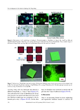
The development of cell-adhesive hydrogel for 3D printing


            Figure 6.  Observation of cell  morphology in hydrogels.  Photomicrographs of  fibroblasts in Alg-gel  (A), Alg-Ph gel  (B), and
            Alg-Ph/Gelatin-Ph gel (C). Fibroblasts were stained with Hoechst 33342 and Alexa Fluor 488 phalloidin. Phase contrast images of
            fibroblasts in Alg-gel (D), Alg-Ph gel (E), and Alg-Ph/Gelatin-Ph gel (F). The scale bar is 100 µm.


            Figure 7. Fabrication of 3D gel sheet structure by using a 3D-bioprinter. Bitmap image of mesh structure (A) and 3D computer mod-
            el (B). The fabricated 3D gel sheet strutrure of Alg-Ph/Gelatin-Ph by using 3D-bioprinter (C). The fabricated gel structure was ob-
            served by using a fluorescence microscope (D).

            including  living  cells was fabricated  and  cultured  at   hand, the fibroblasts were confirmed to extend in the 3D
            different time periods, i.e., 1 day or 7 days (Figures 8–9).   gel sheet after 7 days of cultivation (Figures 9D–9F).
            After cultivation, the F-actin of fibroblasts in the 3D gel
            sheet structure was stained and observed. It was found   4. Discussion
            that fibroblast morphology was maintained in the fa-  There  is  an  increasing  demand  in tissue  engineering
            bricated gels at day 1 (Figures 8D–8F). On the other   and  regenerative medicine  research  to  construct 3D

            158                         International Journal of Bioprinting (2016)–Volume 2, Issue 2