
Page 103 - IJB-2-2
P. 103
Kenichi Arai, Yoshinari Tsukamoto, Hirotoshi Yoshida, et al.
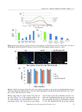
Figure 4. Absorbance spectrum of gelatin, Gelatin-Ph, sodium alginate, and Alg-Ph solution (A). The columns show viscosities of
Alg-Ph (B) and Alg-Ph/Gelatin-Ph (C) solutions. Viscosity of 0.8% sodium alginate solution was used as a control.
Figure 5. Viability test response of fibroblast cells in the hydrogels. Fibroblasts in Alg, Alg-Ph, and Alg-Ph/Gelatin-Ph hydrogels
were stained with calcein-AM and PI at day 1 (A). Cell viability of fibroblasts in Alg-Ph and Alg-Ph/Gelatin-Ph gels at day 1, day 3,
day 5, and day 7 (B). The scale bar is 100 µm.
Bitmap image data of a mesh structure was used in layer by layer to fabricate a 3D lattice structure. As a
this study (Figure 7A). A 3D computer model of the result, the Alg-Ph/Gelatin-Ph 3D gel sheet structure
3D mesh structure was reconstructed from these bit- was successfully fabricated by 3D-bioprinter (Figures
map images (Figure 7B). Twenty layers were printed 7C–7D). The Alg-Ph/Gelatin-Ph 3D gel sheet structure
International Journal of Bioprinting (2016)–Volume 2, Issue 2 157