
Page 102 - IJB-2-2
P. 102
The development of cell-adhesive hydrogel for 3D printing
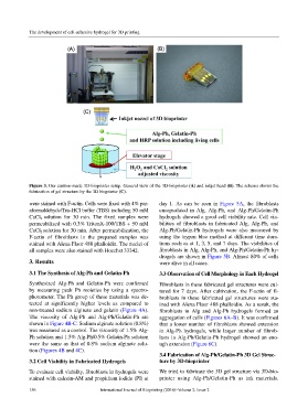
Figure 3. Our custom-made 3D-bioprinter setup. General view of the 3D-bioprinter (A) and inkjet head (B). The schema shows the
fabrication of gel structure by the 3D bioprinter (C).
were stained with F-actin. Cells were fixed with 4% par- day 1. As can be seen in Figure 5A, the fibroblasts
aformaldehyde/Tris-HCl buffer (TBS) including 50 mM encapsulated in Alg, Alg-Ph, and Alg-Ph/Gelatin-Ph
CaCl 2 solution for 30 min. The fixed samples were hydrogels showed a good cell viability rate. Cell via-
permeabilized with 0.3% TritonX-100/TBS + 50 mM bilities of fibroblasts in fabricated Alg, Alg-Ph, and
CaCl 2 solution for 30 min. After permeabilization, the Alg-Ph/Gelatin-Ph hydrogels were also measured by
F-actin of fibroblasts in the prepared samples was using the trypan blue method at different time dura-
stained with Alexa Fluor 488 phalloidin. The nuclei of tions such as at 1, 3, 5, and 7 days. The viabilities of
all samples were also stained with Hoechst 33342. fibroblasts in Alg, Alg-Ph, and Alg-Ph/Gelatin-Ph hy-
drogels are shown in Figure 5B. Almost 80% of cells
3. Results were alive in all cases.
3.1 The Synthesis of Alg-Ph and Gelatin-Ph 3.3 Observation of Cell Morphology in Each Hydrogel
Synthesized Alg-Ph and Gelatin-Ph were confirmed Fibroblasts in these fabricated gel structures were cul-
by measuring peak Ph moieties by using a spectro- tured for 7 days. After cultivation, the F-actin of fi-
photometer. The Ph group of these materials was de- broblasts in these fabricated gel structures were sta-
tected at significantly higher levels as compared to ined with Alexa Fluor 488 phalloidin. As a result, the
non-treated sodium alginate and gelatin (Figure 4A). fibroblasts in Alg and Alg-Ph hydrogels formed an
The viscosity of Alg-Ph and Alg-Ph/Gelatin-Ph are aggregation of cells (Figures 6A–B). It was confirmed
shown in Figure 4B-C. Sodium alginate solution (0.8%) that a lower number of fibroblasts showed extension
was measured as a control. The viscosity of 1.5% Alg- in Alg-Ph hydrogels, while larger number of fibrob-
Ph solution and 1.5% Alg-Ph/0.5% Gelatin-Ph solution lasts in Alg-Ph/Gelatin-Ph hydrogel showed an eno-
were the same as that of 0.8% sodium alginate solu- ugh extension (Figure 6C).
tion (Figures 4B and 4C).
3.4 Fabrication of Alg-Ph/Gelatin-Ph 3D Gel Struc-
3.2 Cell Viability in Fabricated Hydrogels ture by 3D-bioprinter
To evaluate cell viability, fibroblasts in hydrogels were We tried to fabricate the 3D gel structure via 3D-bio-
stained with calcein-AM and propidium iodide (PI) at printer using Alg-Ph/Gelatin-Ph as ink materials.
156 International Journal of Bioprinting (2016)–Volume 2, Issue 2