
Page 101 - IJB-2-2
P. 101
Kenichi Arai, Yoshinari Tsukamoto, Hirotoshi Yoshida, et al.
2.2 The Synthesis of Alginate-Ph and Gelatin-Ph
Molecules
The synthetic methods of producing Alginate-Ph (Alg-
Ph) and Gelatin-Ph were based on previously reported
methods [21–23] . Sodium alginate and gelatin were dis-
solved in MES buffer solution, respectively. These
solutions were activated by using 1-ethyl-3-(3-dim-
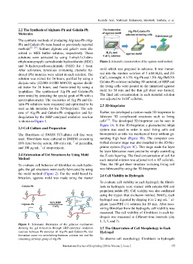
ethylaminopropyl) carbodiimide hydrochloride (EDC) Figure 2. Schematic representation of the agarose mold method.
and N-hydroxysulfosuccinimide (NHS) for 1 hour.
After activation, tyramines containing phenolic hy- mold which was prepared in advance. It was immer-
droxyl (Ph) moieties were added in each solution. The sed into the mixture solution of 5 mM H 2O 2 and 2%
solution was stirred for 24 hours, purified by using a CaCl 2 overnight. A 1.5% Alg-Ph and 1.5% Alg-Ph/0.5%
dialysis tube (12,000–14,000 MWCO) against distill- Gelatin-Ph solution including 50 units/mL of HRP and
ed water for 24 hours, and freeze-dried by using a the living cells were poured in the immersed agarose
lyophilizer. The synthesized Alg-Ph and Gelatin-Ph mold for 30 min and the thin gel sheet was formed.
were tested by detecting the special peak of Ph with a The final cell concentration in each material solution
6
spectrophotometer. The viscosities of Alg-Ph and Ge- was adjusted to 1×10 cells/mL.
latin-Ph solutions were measured and optimized to be 2.5 3D-bioprinter
used as ink materials for the 3D-bioprinter. The sch-
eme of Alg-Ph and Gelatin-Ph conjugation and hy- Earlier, we developed a custom-made 3D-bioprinter to
drogelation by the HRP-catalyzed oxidation reaction fabricate 3D complicated structures such as living
is shown in Figure 1. cells [15] . The developed 3D-bioprinter can be seen in
Figure 3A. In this 3D-bioprinter, a piezoelectric inkjet
2.3 Cell Culture and Preparation system was used in order to eject living cells and
The fibroblasts of SWISS 3T3-albino cell line were biomaterials as inks via mechanical force without ge-
used. Fibroblasts were cultured in DMEM containing nerating high heat (Figure 3B). The computer con-
−1
10% fetal bovine serum, 100 units mL of penicillin, trolled elevator stage was also installed in the 3D-bio-
−1
and 100 µg mL of streptomycin. printer system (Figure 3C). This stage made the layer
by layer fabrication more precise by simply controlling
2.4 Fabrication of Gel Structures by Using Mold the Z-axis layering. The final concentration of cell for
Method each material solution was adjusted to 6 × 10 cells/mL.
6
To evaluate cell behavior of fibroblast in each hydro- Thus, the 3D gel sheet structure including living cell
gels, the gel structures were easily fabricated by using was fabricated by using the 3D-bioprinter.
the mold method (Figure 2). For the mold based fa- 2.6 Cell Viability in Hydrogels
brication, agarose mold was made using the master
To evaluate cell viability in each hydrogel, the fibrob-
lasts in hydrogels were stained with calcein-AM and
propidium iodide (PI). Cell viability was also confirmed
using the trypan blue exclusion method. Briefly, each
−1
hydrogel was digested by dipping it in 2 mg mL al-
ginate lyase/PBS (+) solution for 20 min. After reco-
vering fibroblast from the hydrogels, cell viability was
measured. The cell viability of fibroblasts in each hy-
drogels was measured at different time intervals (day
1, 3, 5, and 7).
Figure 1. Schematic illustration of the gelation mechanism
showing the gel formation through HRP-catalyzed oxidation 2.7 The Observation of Cell Morphology in Each
reaction between Ph moieties of Alg-Ph and Gelatin-Ph. Gel Hydrogel
formation occur via crosslinking between calcium ion and the
remaining carboxyl group of Alg-Ph. To observe cell morphology, fibroblasts in hydrogels
International Journal of Bioprinting (2016)–Volume 2, Issue 2 155