
Page 96 - IJB-2-2
P. 96
Morphological, mechanical and biological assessment of PCL/pristine graphene scaffolds for bone regeneration
that at day 3, all scaffolds exhibited similar biological tion tests of ADSCs were performed by the supplier
performance. At day 7, 0.78% PCL/pristine graphene (ThermoFisher Scientific, UK). The focus of this pa-
scaffolds exhibited greater fluorescence intensity, sta- per was to access the effect of low concentration of
tistically different form 0%, corresponding to a high pristine graphene on both cell viability and prolifera-
cellular activity. This observation can indirectly be tion. Differentiations studies, not reported here, are
correlated to higher cell proliferation rate. On the other being conducted.
hand, based on the statistical analysis, it is possible to 4. Conclusion
notice that on day 14, 0.50% and 0.78% PCL/ pristine
graphene scaffolds positively deviated from PCL sca- This paper presents the morphological, mechanical,
ffolds and 0.13% PCL/pristine graphene scaffolds, and biological properties of PCL/pristine graphene
showing higher cell viability/proliferation rates. It is scaffolds containing different concentrations of pris-
also possible to observe that through all the time po- tine graphene. The results indicate that the screw as-
ints, the fluorescence activity increased, which indi- sisted additive manufacturing system considered in
cates an increase in the cell proliferation rate. The neg- this research work is a viable technique to produce
ative control (NC) shows no metabolically active cells. these composite scaffolds. The results also show that
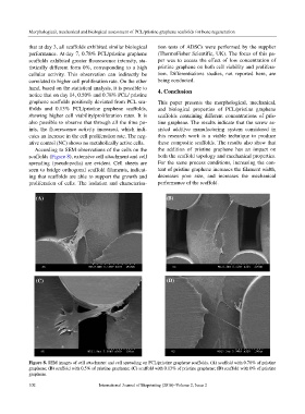
According to SEM observations of the cells on the the addition of pristine graphene has an impact on
scaffolds (Figure 8), extensive cell attachment and cell both the scaffold topology and mechanical properties.
spreading (pseudopodia) are evident. Cell sheets are For the same process conditions, increasing the con-
seen to bridge orthogonal scaffold filaments, indicat- tent of pristine graphene increases the filament width,
ing that scaffolds are able to support the growth and decreases pore size, and increases the mechanical
proliferation of cells. The isolation and characterisa- performance of the scaffold.
(A) (B)
(C) (D)
Figure 8. SEM images of cell attachment and cell spreading on PCL/pristine graphene scaffolds. (A) scaffold with 0.78% of pristine
graphene; (B) scaffold with 0.5% of pristine graphene; (C) scaffold with 0.13% of pristine graphene; (D) scaffold with 0% of pristine
graphene.
102 International Journal of Bioprinting (2016)–Volume 2, Issue 2