
Page 92 - IJB-2-2
P. 92
Morphological, mechanical and biological assessment of PCL/pristine graphene scaffolds for bone regeneration
∆ h
ε = (3)
h 0
where A is the initial sample cross section area and Δh
is the scaffold height variation. The obtained stress-
strain data was further processed to determine the
compression modulus, E c, according to the procedure
[7]
previously reported by Fiedler .
2.7 Biological Test (In Vitro)
Scaffold Preparation
For biological tests, PCL and PCL/pristine graphene
scaffolds were cut into small blocks (11 mm × 11 mm
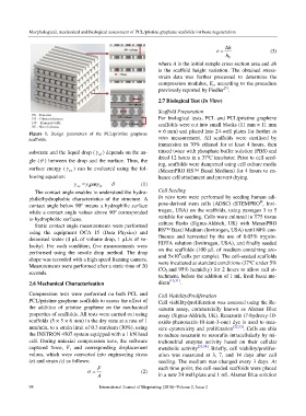
Figure 1. Design parameters of the PCL/pristine graphene × 6 mm) and placed into 24-well plates for further in
scaffolds. vitro measurement. All scaffolds were sterilised by
immersion in 70% ethanol for at least 4 hours, then
substrate and the liquid drop (γ ) depends on the an- rinsed twice with phosphate buffer solution (PBS) and
sl
gle (θ ) between the drop and the surface. Thus, the dried 12 hours in a 37ºC incubator. Prior to cell seed-
ing, scaffolds were dampened using cell culture media
surface energy (γ sv ) can be evaluated using the fol- (MesenPRO RS™ Basal Medium) for 4 hours to en-
lowing equation: hance cell attachment and prevent drying.
γ sv .cosγ = sl γ + lv θ (1)
The contact angle enables to understand the hydro- Cell Seeding
philic/hydrophobic characteristics of the structure. A In vitro tests were performed by seeding human adi-
®
contact angle below 90º means a hydrophilic surface pose-derived stem cells (ADSC) (STEMPRO , Invi-
while a contact angle values above 90º corresponded trogen, USA) on the scaffolds, using passages 3 to 5
to hydrophobic surfaces. suitable for seeding. Cells were cultured in T75 tissue
Static contact angle measurements were performed culture flasks (Sigma-Aldrich, UK) with MesenPRO
using the equipment OCA 15 (Data Physics) and RS™ Basal Medium (Invitrogen, USA) until 80% con-
deionised water (4 µL of volume drop, 1 µL/s of ve- fluence and harvested by the use of 0.05% trypsin-
locity). For each condition, five measurements were EDTA solution (Invitrogen, USA), and finally seeded
performed using the sessile drop method. The drop on the scaffolds (100 µL of medium containing aro-
4
shape was recorded with a high speed framing camera. und 5×10 cells per sample). The cell-seeded scaffolds
Measurements were performed after a static time of 20 were incubated at standard conditions (37ºC under 5%
CO 2 and 95% humidity) for 2 hours to allow cell at-
seconds.
tachment, before the addition of 1 mL fresh basal me-
2.6 Mechanical Characterisation dium [16,31] .
Compression tests were performed on both PCL and Cell Viability/Proliferation
PCL/pristine graphene scaffolds to assess the effect of Cell viability/proliferation was assessed using the Re-
the addition of pristine graphene on the mechanical sazurin assay, commercially known as Alamar Blue
properties of scaffolds. All tests were carried out using assay (Sigma-Aldrich, UK). Resazurin (7-hydroxy-10-
scaffolds (5 × 5 × 6 mm) in the dry state at a rate of 1 oxido-phenoxazin-10-ium-3-one) dye is used to mea-
mm/min, to a strain limit of 0.3 mm/mm (30%), using sure cytotoxicity and proliferation [32,33] . Cells are able
the INSTRON 4507 system equipped with a 1 kN load to reduce resazurin to resorufin intracellularly by mi-
cell. During uniaxial compression tests, the software tochondrial enzyme activity based on their cellular
captured force, F, and corresponding displacement metabolic activity [33,34] . Briefly, cell viability/prolifer-
values, which were converted into engineering stress ation was measured at 3, 7, and 14 days after cell
(σ) and strain (ε) as follows: seeding. The medium was changed every 3 days. At
F each time point, the cell-seeded scaffolds were placed
σ = (2)
A in a new 24 well plate and 1 mL Alamar Blue solution
98 International Journal of Bioprinting (2016)–Volume 2, Issue 2