
Page 67 - IJB-2-2
P. 67
Atra Malayeri, Colin Sherborne, Thomas Paterson, et al.
approach, the inherent porosity dictated by the emul- tissue engineering constructs. A human osteosarcoma
sion templating is retained, while the larger pore sizes cell line (MG63) was cultured on bulk and woodpile
(to enhance materials transport and ingrowth) are built PolyHIPE for up to 7 days as well as tissue culture
by laser-based direct write. plastic as positive control. Figure 3 and Figure 4 show
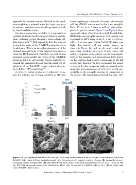
For tissue engineering scaffolds, it is important to successful culture of MG63 cells on both EHA80 Poly-
consider additional features beyond structural archite- HIPE disks and woodpile structures. Cell viability was
cture, including surface chemistry which affects cel- evaluated via MTT assay on day 1, 3 and 7. Cell via-
lular attachment [15] . With regards to this, the inherent bility of acrylic acid coated PolyHIPE disks was
hydrophobic nature of the PolyHIPE material needs to higher than control in all time points. However, as
be addressed. This is an inevitable consequence of the shown in Figure 4A both acrylic acid coated and
required hydrophobicity of the original monomers to non-coated woodpile structures showed lower cell
create the HIPE emulsion. Therefore, we used plasma activity compared to the control, but the biocompati-
treatment to post-modify the surface of the PolyHIPE bility of the materials was demonstrated. Cells grown
structures prior to cell culture. Plasma treatment in- on the scaffolds had a larger surface area to the 2D
creased the hydrophilicity, and thus the initial cell at- counterpart; therefore we have normalised the assays
tachment of the PolyHIPE surface without affecting to account for this. A separate study was carried out to
the bulk PolyHIPE morphology [22] . determine what percentages of cells were seeded suc-
In vitro cell culture studies were undertaken to as- cessfully on the woodpile structure in comparison to
sess the potential use of these scaffolds as 3D bone the control. The investigation showed that only 36%
Figure 3. In vitro experiment with PolyHIPE disks. (A) MTT assay for proliferation of MG63 on acrylic acid coated and non-coated
PolyHIPE disks during different incubation period. Error bars represent the standard deviation of mean. (B) Immunofluorescence
micrograph of cryo-sectioned PolyHIPE disk cultured with MG63 stained with DAPI and Phalloidin-FITC at day 7 – Scale bar 100
μm. (C) SEM micrographs showing the attachment of MG63 cells on non-coated PolyHIPE disks on day 7, (D) on acrylic acid
coated PolyHIPE disks on day 7.
International Journal of Bioprinting (2016)–Volume 2, Issue 2 73