
Page 65 - IJB-2-2
P. 65
Atra Malayeri, Colin Sherborne, Thomas Paterson, et al.
Phalloidin was excited using a 488 nm laser (4% tran- example a closed surface is observed when the HIPE
[2]
smission) and emission detected above 505 nm. For is cured against polypropylene . Similar features
differential interference contrast (DIC), a 543 nm laser were observed in the HIPE emulsion when selectively
(21.8% transmission) was used without filters to pro- curing regions within the bulk emulsion. The boun-
duce a contrast of scaffold. Z-stacks were converted to dary between the cured and uncured HIPE formed a
single images using ImageJ’s Z-project feature using surface skin during the post-processing stages to wash
the max intensity blend setting. Images were measured and dry the PolyHIPE structures. This is attributed to a
and scale bars were also added by ImageJ. structural collapse of a partially cured boundary layer
on the surface of the PolyHIPE. There are two plausi-
3. Results and Discussion ble explanations for this phenomenon: (i) the HIPE
3.1 Morphological Characterisation material acted as a scattering medium for the curing
UV light, and (ii) the diffusion of reactive radicals
The morphology and surface structure of the woodpile from the photo-initiated region.
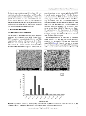
structures were analysed using SEM. Typical Poly- The microporosity can be controlled by the speed
HIPE morphology was maintained within the scaffolds, of the paddle stirrer. The pore size of the polyHIPE
suggesting the selective polymerisation of the Poly- produced at a stir speed of 350 rpm was determined
HIPE did not adversely affect the internal porosity. using SEM (Figure 1). ImageJ was used to measure
The creation of a surface skin has been reported in the pore diameters from fractured PolyHIPE structures
literature when the HIPE collapses at the surface, for and to account for the underestimation of the measured
Figure 1. PolyHIPE disk morphology. (A) Morphology of 80% EHA80 PolyHIPE disks obtained by SEM – Scale bar 100 μm, (B)
Scale bar 50 μm. (C) Void diameter distribution of PolyHIPE disks based on SEM micrograph analysis.
International Journal of Bioprinting (2016)–Volume 2, Issue 2 71