
Page 280 - IJB-10-2
P. 280
International Journal of Bioprinting Microfluidic spinning for neural models
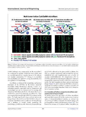
Figure 3. Perfusion fluid strategy and characterization of multi-lumen hollow CaA/GelMA composite microfibers. (A) CaA/GelMA double-lumen
microfiber with one kind of inclusion. (B) CaA/GelMA double-lumen microfiber with two kinds of inclusions. (C) CaA/GelMA triple-lumen microfiber
with two kinds of inclusions. Scale bar = 100 µm.
GelMA hydrogel was compounded in the microfiber. of HUVECs cultured on the plates tended to flatten. The
48
As a substitute for gelatin, GelMA has been widely used vWF is a protein synthesized and secreted by vascular
as a scaffold material for vascular tissues in 3D cultures endothelial cells and megakaryocytes and a marker of
and vascular construction. 33,48,49 Owing to its excellent vascular endothelialization. We characterized vWF in
biocompatibility and rapid UV curing processability, it is HUVECs cultured in composite microfibers for 7 days and
also applied in 3D bioprinting. compared the expression of vWF in HUVECs cultured on
a plate. The high-magnification laser confocal microscopy
In addition, we observed the morphology and protein
expression of the HUVECs in the hollow composite images (Figure 4C and E) show that HUVECs cultured in
composite microfibers exhibited a positive expression of
microfibers using fluorescence staining. F-actin is a vWF, which was brighter than that in HUVECs cultured in
cytoskeletal protein commonly used to characterize cell two-dimensional culture.
morphology and can be characterized by staining with
Alexa Fluor 488-labeled phalloidin. We stained the F-actin 3.5. Assembly of hollow composite microfibers and
of HUVECs cultured in composite microfibers for 7 days construction of neural model
and compared the morphology of HUVECs cultured on the To further expand the application of hollow microfibers
plates. The high-magnification laser confocal microscopy and make them convenient for the construction of various
images (Figure 4B and D) reveal that HUVECs cultured in types of disease/physiological models, we realized the
composite microfibers have an enhanced 3D morphology 3D printing of hollow composite microfibers using a 3D
and a tendency to extend, closely resembling the printing platform built in the laboratory (Figure 1B). The
41
morphology of real living cells. Moreover, the morphology fluorescence images of 3D-printed hollow microfiber are
Volume 10 Issue 2 (2024) 272 doi: 10.36922/ijb.1797