
Page 291 - IJB-10-2
P. 291
International Journal of Bioprinting Kidney hydrogel print for renal cancer model
24-well plates, cultured for 7 days, and the medium was to the product’s protocol. Real-time polymerase chain
changed every other day. reaction (RT-PCR; Roche, Switzerland) was used to analyze
the relative gene expression. To perform RT-PCR, cDNA
2.17. Cellular proliferation measurement (50 ng), specific primers (Table 1), and SYBR Master Mix
The effect of porcine kidney-derived dECM on ACHN cell were mixed to a total reaction volume of 10 µL.
proliferation was analyzed using CCK-8 assays on days 1, 3,
5, and 7 for all bioprinted nephron models. Samples from all 2.21. RNA sequencing and data analysis
groups were transferred to new culture plates and incubated Transcriptome sequencing and analysis were conducted
with CCK-8 working solution (10% v/v) that dissolved with the by OE Biotech Co., Ltd. (China). The RNA integrity was
fully supplemented DMEM for 1.5 h. Finally, the absorbance assessed using an Agilent 2100 Bioanalyzer (Agilent
of the incubated medium was read at 450 nm. The cellular Technologies, USA). Libraries were sequenced on an
proliferation rate at each time point was then determined Illumina NovaSeq 6000 platform, and 150 bp paired-end
using the absorbance value of day 1 as the baseline. reads were generated, with approximately 50 raw reads
generated for each sample. Principal component analysis
2.18. Cellular morphology staining was performed using R version 3.2.0 to evaluate the
The cellular morphology of ACHN cells in all bioprinted biological duplication of samples. Differential expression
nephron models was fixed with 4% PFA and stained with analysis was performed using DESeq2. P value <0.05 and
phalloidin on day 5. Samples were incubated with 0.5% Log2FC >1 or Log2FC <-1 were set as the thresholds
Triton X-100 for 15 min, phalloidin working solution to identify significantly differentially expressed genes
(1:200) for 40 min, and DAPI solution for 10 min in the (DEGs). Gene Ontology (GO) pathway enrichment
dark. Finally, the samples were washed with PBS and analysis of DEGs was performed to screen for significantly
imaged (n = 6 images at different spots per group) using a enriched terms. Gene set enrichment analysis (GSEA) was
confocal microscopy. performed using GSEA software.
2.19. RNA extraction 2.22. Immunofluorescence staining
Total RNA samples from all bioprinted nephron models All bioprinted nephron models were fixed on day 5
and casted models were lysed with TRIzol, followed by using 4% PFA at room temperature, washed in PBS
mixing with chloroform, washing with isopropanol and three times, permeabilized with 0.2% Triton X-100 for
ethanol, and dissolved with DEPC water, and the final 30 min, and blocked with 5% bovine serum albumin for
RNA concentration was determined with Nanodrop™ 2000 1 h. Primary antibodies (1:200) targeting CD44, TWIST,
Spectrophotometer (Thermo Fisher Scientific, USA). vimentin, CDH2, and CDH1 were added to the samples
and incubated at room temperature for 2 h. Afterward,
2.20. Real-time quantitative polymerase chain the samples was washed with PBS thrice, and then stained
reaction with secondary antibodies (1:200) for 1 h and DAPI for 5
The complementary DNA (cDNA) was reverse-transcribed min. Secondary antibody assay was also performed, where
with a HiScript III All-in-one RT SuperMix kit, according secondary antibody was directly stained after blocking,
®
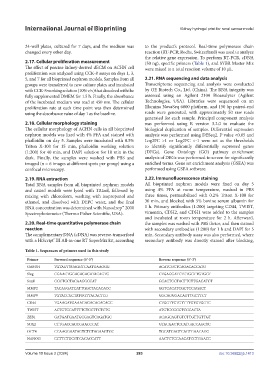
Table 1. Sequences of primers used in this study
Primer Forward sequence (5’-3’) Reverse sequence (5’-3’)
GAPDH TGTAGTTGAGGTCAATGAAGGG ACATCGCTCAGACACCATG
Slug CGAACTGGACACACATACAGTG CTGAGGATCTCTGGTTGTGGT
Snail GGCTGCTACAAGGCCAT GCACTGGTACTTCTTGACATCT
MMP2 TACAGGATCATTGGCTACACACC GGTCACATCGCTCCAGACT
MMP9 TGTACCGCTATGGTTACACTCG GGCAGGGACAGTTGCTTCT
CD44 TGAAGATGAAAGAGACAGACACC CTGGTTCTGTTTTGTGTGGTC
TWIST ACTGTCCATTTTCTCCTTCTCTG ATGTCCGCGTCCCACTA
ZEB1 GATGATGAATGCGAGTCAGATGC ACAGCAGTGTCTTGTTGTTGT
SOX2 CTTGACCACCGAACCCAT GTACAACTCCATGACCAGCTC
OCT4 CCAAGGAATAGTCTGTAGAAGTGC TGCATGAGTCAGTGAACAGG
NANOG CCTTCTGCGTCACACCATT AACTCTCCAACATCCTGAACC
Volume 10 Issue 2 (2024) 283 doi: 10.36922/ijb.1413