
Page 293 - IJB-10-2
P. 293
International Journal of Bioprinting Kidney hydrogel print for renal cancer model
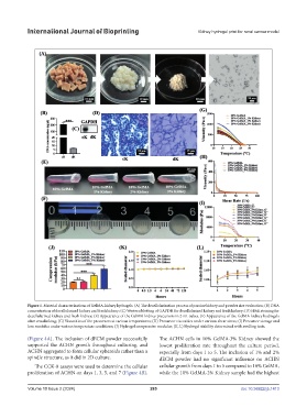
Figure 1. Material characterizations of GelMA-kidney hydrogels. (A) The decellularization process of porcine kidney and powder size evaluation; (B) DNA
concentration of decellularized kidney and fresh kidney; (C) Western blotting of GAPDH for decellularized kidney and fresh kidney; (D) H&E staining for
decellularized kidney and fresh kidney; (E) Appearance of the GelMA-kidney precursors in 2 mL tubes. (F) Appearance of the GelMA-kidney hydrogels
after crosslinking; (G) Viscosities of the precursors at various temperatures; (H) Precursor viscosities under various shear stress; (I) Precursor storage and
loss modulus under various temperature conditions; (J) Hydrogel compressive modulus; (K, L) Hydrogel stability determined with swelling tests.
(Figure 4A). The inclusion of dECM powder successfully The ACHN cells in 10% GelMA-3% Kidney showed the
supported the ACHN growth throughout culturing, and lowest proliferation rate throughout the culture period,
ACHN aggregated to form cellular spheroids rather than a especially from days 1 to 5. The inclusion of 1% and 2%
spindle structure, as it did in 2D culture. dECM powder had no significant influence on ACHN
The CCK-8 assays were used to determine the cellular cellular growth from days 1 to 5 compared to 10% GelMA,
proliferation of ACHN on days 1, 3, 5, and 7 (Figure 4B). while the 10% GelMA-2% Kidney sample had the highest
Volume 10 Issue 2 (2024) 285 doi: 10.36922/ijb.1413