
Page 380 - IJB-10-2
P. 380
International Journal of Bioprinting AM evaluation of medical device companies
Table 4. Continued...
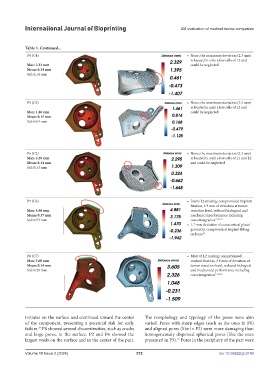
P4 (C4) • None; the maximum deviation (2.3 mm)
is located in only a few cells of L2 and
Max: 2.33 mm could be neglected
Mean: 0.14 mm
Std: 0.14 mm
P5 (C5) • None; the maximum deviation (1.5 mm)
is located in only a few cells of L3 and
Max: 1.46 mm could be neglected
Mean: 0.15 mm
Std: 0.63 mm
P6 (C2) • None; the maximum deviation (2.3 mm)
Max: 2.29 mm is located in only a few cells of L1 and L2
Mean: 0.14 mm and could be neglected
Std: 0.13 mm
P7 (C6) • Entire L2 missing; compromised implant
fixation, 4.9 mm of deviation at tumor
Max: 4.88 mm resection level, reduced biological and
Mean: 0.37 mm mechanical performance including
Std: 0.53 mm osseointegration 15,22,23
• 1.7 mm deviation of extracortical plates’
geometry, compromised implant fitting
on bone 15
P8 (C7) • Most of L2 missing; compromised
Max: 3.60 mm implant fixation, 3.6 mm of deviation at
Mean: 0.14 mm tumor resection level, reduced biological
Std: 0.28 mm and mechanical performance including
osseointegration 15,22,23
initiates on the surface and continued toward the center The morphology and typology of the pores were also
of the component, presenting a potential risk for early varied. Pores with sharp edges (such as the ones in P8)
failure. P8 showed several discontinuities, such as cracks and aligned pores (like in P2) were more damaging than
24
and large pores, in the surface. P2 and P6 showed the homogeneously dispersed spherical pores (like the ones
largest voids on the surface and in the center of the part. presented in P3). Pores in the periphery of the part were
25
Volume 10 Issue 2 (2024) 372 doi: 10.36922/ijb.0140