
Page 486 - IJB-10-2
P. 486
International Journal of Bioprinting Bioprinted skin for testing of therapeutics
proof-of-concept that human skin equivalents produced, serum (FBS; Thermo Fisher Scientific, UK). The dermal
using scalable, automated biofabrication techniques can samples were incubated using standard culture conditions
be used as a preclinical tool to predict potential adverse (37 C with 5% CO ) for 24 h. After 24 h, wells were topped
o
2
immune responses of therapeutic antibodies. up with 5 mL of Media A (Table 1). Dermal dissects were
observed for fibroblast outgrowth with media changed
2. Materials and methods twice weekly. Once significant outgrowth was observed,
fibroblasts were dissociated using trypsin/EDTA (Sigma-
2.1. Cell culture Aldrich) using standard culture conditions for 5 min. The
Full Local Research Ethics Committee (LREC) approval dissociated fibroblasts were further cultured for up to 7
was obtained before sourcing healthy volunteer samples passages. Keratinocytes were dissociated from epidermal
for this study. After obtaining informed consent, two 4 samples using trypsin/EDTA using standard cell culture
mm skin biopsies and 60 mL of whole blood were collected conditions. The resulting cells were further cultured in
from healthy volunteers. Skin biopsies were rinsed in Media B (Table 1) for up to 3 passages with media changed
phosphate-buffered saline (PBS; Sigma-Aldrich, UK), twice weekly. Lymphoprep™ density gradient medium
then excess adipose tissue was removed from the skin (STEMCELL Technologies, UK) was used to isolate human
biopsies, and the biopsies were incubated in 1.0 U/mL peripheral blood monocytes (PBMCs) from whole blood
dispase solution (Scientific Laboratory Supplies, UK) for sourced from healthy volunteers as previously described. 8
18 h at 4°C to allow separation of the epidermis from the
dermis. Once separated, the dermis and epidermis were 2.2. Bioprinting equipment and process
processed independently. To isolate dermal fibroblasts, For this study, a Microfab Jetlab 4 XL (Microfab Technologies
the separated dermal samples were dissected, placed into Inc., US) printer was retrofitted to accommodate
6-well plates, and covered with 100 µL of fetal bovine solenoid VHS series microvalves (Lee Products Ltd., US)
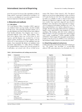
Table 1. Media formulations and working concentrations
Media Components Supplier Final concentration
A Dulbecco’s Modified Eagles Medium Sigma-Aldrich, UK -
Fetal calf serum Fisher Scientific, UK 10%
Penicillin/Streptomycin/Amphotericin Sigma-Aldrich, UK 1%
L-glutamine Sigma-Aldrich, UK 1%
B Epilife Fisher Scientific, UK -
Human Keratinocyte Growth Supplement Fisher Scientific, UK 1%
Penicillin/Streptomycin/Amphotericin Sigma-Aldrich, UK 1%
L-glutamine Sigma-Aldrich, UK 1%
C 3 Part Dulbecco’s Modified Eagles Medium Sigma-Aldrich, UK -
1 Part Ham’s F12 Nutrient Mixture Sigma-Aldrich, UK -
Chelex-treated fetal calf serum Fisher Scientific, UK 5%
Penicillin/Streptomycin/Amphotericin Sigma-Aldrich, UK 1%
L-glutamine Sigma-Aldrich, UK 1%
Cholera toxin Sigma-Aldrich, UK 8.5 ng/mL
Hydrocortisone Sigma-Aldrich, UK 0.4 μg/mL
Human insulin Sigma-Aldrich, UK 5 μg/mL
Adenine Sigma-Aldrich, UK 24 μg/mL
Human epidermal growth factor PeproTech, US 20 ng/mL
D 3 Part Dulbecco’s Modified Eagles Medium Sigma-Aldrich, UK -
1 Part Ham’s F12 Nutrient Mixture Sigma-Aldrich, UK -
Fetal calf serum Fisher Scientific, UK 10%
Penicillin/Streptomycin/Amphotericin Sigma-Aldrich, UK 1%
Cholera toxin Sigma-Aldrich, UK 8.5 ng/mL
Hydrocortisone Sigma-Aldrich, UK 0.4 μg/mL
Human epidermal growth factor PeproTech, US 20 ng/mL
Human transferrin PeproTech, US 5 μg/mL
L-ascorbic acid Sigma-Aldrich, UK 100 μg/mL
F RPMI-1640 Sigma-Aldrich, UK -
Autologous serum - 20%
Penicillin/Streptomycin/Amphotericin Sigma-Aldrich, UK 1%
Volume 10 Issue 2 (2024) 478 doi: 10.36922/ijb.1851