
Page 487 - IJB-10-2
P. 487
International Journal of Bioprinting Bioprinted skin for testing of therapeutics
to create a high-throughput bioprinting platform. The the bioink. The magnet within the reservoir was rotated
inkjet printhead of the Jetlab 4 printer was replaced by an additional rotating magnet located perpendicular
with a microvalve printhead (Figure 1), which could to the external wall of the reservoir. The external magnet
accommodate four individual custom-built ink reservoirs, was connected to a 24 V DC motor via drive belt, which
each with an ink capacity of 2.5 mL. Inkjet printing is was operated at 18 V for printing of cells. To measure the
very effective for small volumes and can offer single-cell volume dispensed, 200 droplets of media D (Table 1) were
resolution, but the development of skin-equivalent models printed into 1.5 mL Eppendorf tubes (Fisher Scientific),
requires higher volume deposition rates, and so, for this which was weighed using a microbalance (Mettler Toledo).
reason, microvalve printing was preferred. The outlets The volume per droplet was calculated from this data. Dwell
of the reservoirs were threaded to contain a male–male times of 100–1000 µs and backpressures of 50–500 mmHg
062 MINSTAC threaded connector (Lee Products Ltd.); were investigated at increments of 50 µs and 50 mmHg. To
one side of which was fixed into the reservoir (Figure 1). further ensure consistency of output, a microvalve purge
The other end of the male–male connector was used to step was used between print events.
attach the solenoid VHS series. The valve configuration
used in this study had female 062 MINSTAC threaded 2.3. Cell viability post printing
To ensure that the correct number of dermal fibroblasts
inlets and outlets and was used alongside 062 MINSTAC and epidermal keratinocytes were dispensed when printing
jeweled orifice nozzles (Lee Products Ltd., US) with orifice skin equivalents, fibroblast suspensions were prepared
diameters of 190 µm. To generate actuation signals for at a concentration of 25 × 10 cells/mL in Media A and
6
the microvalves, spike and hold electrical drivers (Lee keratinocytes were prepared at a concentration of 20 × 10
6
Products Ltd.) were connected to both the Jetlab 4 device cells/mL in Media B and loaded into reservoirs. Following
signal output channels and the signal connections of the a 60 µL purge of the bioink through the microvalves, 20 µL
Individual valves. This allowed signals produced by the of each cell suspension was printed in three separate wells
waveform printed circuit board (PCB) of the Jetlab to be of a 96-well plate. Cells were printed with a backpressure
converted to a 24 V spike voltage and a 5 V dwell voltage. of 150 mmHg and a dwell time of 1000 µs. As described
The optimal parameters for reliable microdispensing above, a 10 µL aliquot of each printed suspension was then
of low viscosity inks were investigated by exploring the mixed 1:1 with trypan blue, and cell number and viability
impact on adjusting the operating dwell time (time that were quantified using a hemocytometer.
the microvalve remains open during one actuation cycle)
and pneumatic backpressure used to eject droplets from 2.4. Bioprinting of cells for the development of
the reservoir. The custom-designed reservoir housed a autologous skin equivalents
®
gold-plated neodymium magnet, which was suspended Figure 2 illustrates the 96-well Alvetex (Reprocell, UK)
within the bioink and was used to agitate cells within culture system, and the general approach to developing
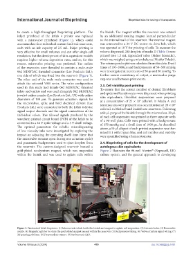
Figure 1. Customized Jetlab bioprinter. (1) Ink reservoir which holds the bioink and magnet to agitate cell suspension. (2) Solenoid valve. (3) Removable
nozzle. (4) Magnetic agitator to rotate the gold-plated magnet present within the reservoir. (5) Backpressure tubing. (6) Valve actuation signal wiring. (7)
XY printing platform. (8) Drop analysis camera. (9) Stroboscopic LED.
Volume 10 Issue 2 (2024) 479 doi: 10.36922/ijb.1851