
Page 490 - IJB-10-2
P. 490
International Journal of Bioprinting Bioprinted skin for testing of therapeutics
successfully deposit material, and attempting to print with dermis of the full-thickness human skin equivalent (HSE)
parameters below these values did not yield successful was well populated with a layer of fibroblasts on top of the
droplet deposition. At lower dwell times (Figure 3A–D), scaffold supporting the epidermis. The epidermis of the
a variance in the volume per droplet was demonstrated HSE contained visible basal, spinous, and granular layers
across the range of backpressures tested. Higher dwell and a very thin stratum corneum. The differentiating and
times (Figure 3E–I) resulted in a more linear increase superficially migrating keratinocytes formed a spinous
in the volume per droplet dispensed across the pressure layer. The layer of granular cells indicated by the darker
range. Peak output was 210 ± 6 nL per droplet, produced at and slightly speckled hematoxylin-stained nuclei (which
a dwell time of 1000 μs with a backpressure of 500 mmHg. separated during sectioning) was located above the spinous
A dwell time of 1000 μs and a backpressure of 150 mmHg keratinocytes. Picrosirius red staining of bioprinted full-
with a volume per droplet of 103 ± 1 nL were used to print thickness skin equivalents showed that the dermis was
cells and produce skin equivalents, giving good balance heavily loaded with collagen (Figure 5C). Large thick
between productivity and consistency of output. crosslinked collagen layers could be seen directly beneath
To print the skin-equivalent models, fibroblasts the epidermis at the dermal–epidermal junction. This layer
were suspended at 25 × 10 cells/mL and keratinocytes of collagen could be seen across the dermis, supporting the
6
were suspended at 20 × 10 cells/mL with 20 µL of each formation of an epidermis.
6
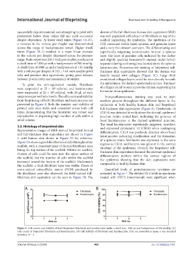
suspension per well into 3 wells. The cell counts and viability Immunofluorescence staining was used to stain
from bioprinting of both fibroblasts and keratinocytes are markers present throughout the different layers in the
presented in Figure 4. Both the number and viability of epidermis of both healthy human skin and bioprinted
printed cells were stable and consistent across both cell full-thickness skin equivalents (Figure 6). Cytokeratin 14
types, demonstrating that the bioprinter was robust and (CK14) was detected directly above the dermal–epidermal
reproducible in dispensing high number of cells within a junction (white dotted line), indicating the presence of
small volume. basal keratinocytes at the dermal epidermal junction.
The basal keratinocytes superficially migrated, stratified,
3.2. Histology of bioprinted skin and expressed cytokeratin 10 (CK10) when undergoing
Representative images of H&E-stained bioprinted dermal differentiation. CK10 was positively detected above basal
and full-thickness skin equivalents are shown in Figure keratinocytes indicating stratification and the formation
5, with human skin shown in Figure 5D for reference. of a spinous strata. Involucrin was expressed in the same
Figure 5A shows a good distribution of cells throughout the regions as CK10, and loricrin was present in the corneal
scaffold, with a consistent layer of dermal fibroblasts seen
lining the top surface of the scaffold. Within the scaffold, envelope of the epidermis. Overall, the bioprinted full-
clusters of cells could be seen near the upper surface of thickness skin equivalents featured the relevant epidermal
the scaffold, but the number of cells within the scaffold differentiation markers within the correct regions of
the epidermis showing that the skin equivalents were
increased toward the bottom of the scaffold. Underneath comparable to healthy human skin.
the scaffold, a thick fibroblast layer was visible. Fibers of
eosin-stained extracellular matrix (ECM) produced by Quantified levels of proinflammatory cytokines are
the fibroblasts were also observed. An H&E-stained full- presented in Figure 7. The elevated IL-2 levels in specimens
thickness skin equivalent can be seen in Figure 5B. The treated with OKT3 (muromonab) were significant when
Figure 4. Cell counts and viability of both bioprinted fibroblasts and keratinocytes under a dwell time 1000 μs and backpressure of 150 mmHg. (A)
Cell counts of bioprinted fibroblasts and keratinocytes. (B) Cell viability of fibroblasts and keratinocytes. Data are presented as mean ± one standard
deviation. N = 3.
Volume 10 Issue 2 (2024) 482 doi: 10.36922/ijb.1851