
Page 499 - IJB-10-2
P. 499
International Journal of Bioprinting Mineralization of 3D-printed PHA scaffolds
2.5. Mechanical properties with ethidium homodimer (red). The cells on the scaffolds
Compression test was evaluated on a universal testing were stained for 1 h in an incubator. Cell proliferation
machine (RB301 UNITECH-M; RnD, Korea) with a 500 was evaluated using the WST-1 assay (Premix WST-1 Cell
kN load cells and a 5 mm/min crosshead speed. The sample Proliferation Assay System; Takara Bio, Shiga, Japan). The
size was 5 × 5 × 3 mm, and the strand size was 400 μm. cells on the scaffolds were immersed in WST-1 solution
2.5. Cell culture for 30 min, and the supernatant was measured at 440 nm
MG63, which is an osteoblast-like cell line, was cultured with a microplate reader (SpectraMax iD3; Molecular
in Dulbecco’s modified Eagle’s medium (DMEM; Gibco/ Devices, San Jose, CA, USA). A radioimmunoprecipitation
Thermo Fisher Scientific, Waltham, MA, USA) with 10% assay (RIPA) buffer (Pierce RIPA buffer; Thermo Fisher
fetal bovine serum and 1% penicillin (Gibco). The cells Scientific) was used to lyse the cells of the scaffold for
were cultured at 37℃ under 5% CO conditions. alkaline phosphatase (ALP) extraction, and the cells were
2
preserved at -80°C. The ALP kit was utilized according to
2.6. Cell viability, proliferation, and differentiation the manufacturer’s instructions. Picogreen dsDNA assay
on a functionalized PHA biopolymer scaffold (Quant-iT™ PicoGreen™ dsDNA Assay Kit; Invitrogen/
The cells were seeded on the PHA biopolymer scaffolds, and Thermo Fisher Scientific, Waltham, MA, USA) was used to
the scaffolds were evaluated for cell survival, proliferation, normalize the ALP activity.
and differentiation. The scaffold was sterilized for 1 min
using 70% ethanol, followed by a phosphate-buffered 2.7. Statistical analysis
saline wash. MG63 cells (5 × 10 cells/mL) were seeded All quantitative data are presented as mean ± standard
5
on each sample and cultured for 7 days for viability and deviation. One-way analysis of variance (ANOVA) was
proliferation, and for 10 days for differentiation. used to analyze the results, followed by Tukey’s post-hoc
Viability was investigated using a live/dead assay kit test. Asterisks in the figures denote significant values (p
(Live and Dead Kit, Invitrogen, USA) containing calcein < 0.05). Statistical analysis was performed using Origin
AM and ethidium homodimer-1. Live cells were stained software (ver. 8.6; OriginLab Corporation, Northampton,
with calcium AM (green), and dead cells were labeled MA, USA).
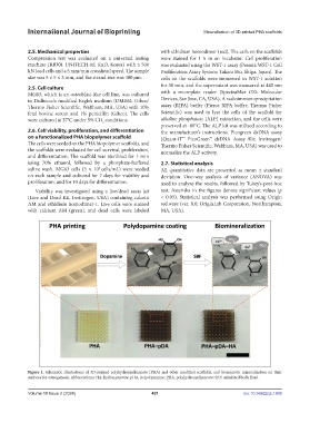
Figure 1. Schematic illustrations of 3D-printed polyhydroxyalkanoate (PHA) and other modified scaffolds, and biomimetic mineralization on their
surfaces for osteogenesis. Abbreviations: HA, hydroxyapatite; pDA, polydopamine; PHA, polyhydroxyalkanoate; SBF: simulated body fluid.
Volume 10 Issue 2 (2024) 491 doi: 10.36922/ijb.1806