
Page 501 - IJB-10-2
P. 501
International Journal of Bioprinting Mineralization of 3D-printed PHA scaffolds
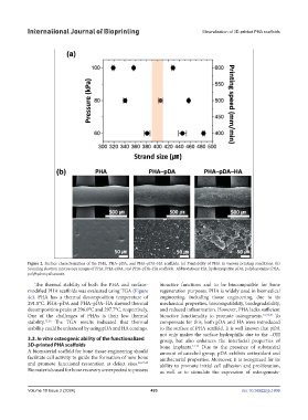
Figure 2. Surface characterization of the PHA, PHA–pDA, and PHA–pDA–HA scaffolds. (a) Printability of PHA in various printing conditions. (b)
Scanning electron microscopy images of PHA, PHA–pDA, and PHA–pDA–HA scaffolds. Abbreviations: HA, hydroxyapatite; pDA, polydopamine; PHA,
polyhydroxyalkanoate.
The thermal stability of both the PHA and surface- bioactive functions and to be biocompatible for bone
modified PHA scaffolds was evaluated using TGA (Figure regeneration purposes. PHA is widely used in biomedical
4e). PHA has a thermal decomposition temperature of engineering, including tissue engineering, due to its
291.8°C. PHA–pDA and PHA–pDA–HA showed thermal mechanical properties, biocompatibility, biodegradability,
decomposition peaks at 296.0°C and 297.7°C, respectively. and reduced inflammation. However, PHA lacks sufficient
One of the challenges of PHAs is their low thermal bioactive functionality to promote osteogenesis. 22,29,30 To
stability. 12,26 The TGA results indicated that thermal compensate for this, both pDA and HA were introduced
stability could be enhanced by using pDA and HA coatings. to the surface of PHA scaffold. It is well known that pDA
not only makes the surface hydrophilic due to the –OH
3.3. In vitro osteogenic ability of the functionalized group, but also enhances the interfacial properties of
3D-printed PHA scaffolds bone implants. 31-33 Due to the presence of substantial
A biomaterial scaffold for bone tissue engineering should amount of catechol group, pDA exhibits antioxidant and
facilitate cell activity to guide the formation of new bone antibacterial properties. Moreover, it is recognized for its
and promote functional restoration at defect sites. 22,27,28 ability to promote initial cell adhesion and proliferation,
Biomaterials used for bone recovery are expected to possess as well as to stimulate the expression of osteogenesis-
Volume 10 Issue 2 (2024) 493 doi: 10.36922/ijb.1806