
Page 505 - IJB-10-2
P. 505
International Journal of Bioprinting Mineralization of 3D-printed PHA scaffolds
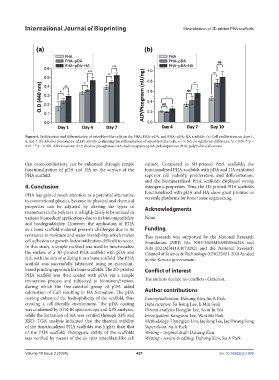
Figure 6. Proliferation and differentiation of osteoblast-like cells on the PHA, PHA–pDA, and PHA–pDA–HA scaffolds. (a) Cell proliferation on days 1,
4, and 7. (b) Alkaline phosphatase (ALP) activity confirming the differentiation of osteoblast-like cells. n = 5; NS, no significant difference; *p < 0.05; **p <
0.01; ***p < 0.001. Abbreviations: ALP, alkaline phosphatase; HA, hydroxyapatite; pDA, polydopamine; PHA, polyhydroxyalkanoate.
that osteoconductivity can be enhanced through simple culture. Compared to 3D-printed PHA scaffolds, the
functionalization of pDA and HA on the surface of the functionalized PHA scaffolds with pDA and HA exhibited
PHA scaffold. superior cell viability, proliferation, and differentiation,
and the biomineralized PHA scaffolds displayed strong
4. Conclusion osteogenic properties. Thus, the 3D-printed PHA scaffolds
functionalized with pDA and HA show great promise as
PHA has gained much attention as a potential alternative versatile platforms for bone tissue engineering.
to conventional plastics, because its physical and chemical
properties can be adjusted by altering the types of Acknowledgments
monomers in the polymer. It is highly likely to be utilized in
various biomedical applications due to its biocompatibility None.
and biodegradability. However, the application of PHA
as a bone scaffold material presents challenges due to its Funding
resistance to moisture and water insolubility, which makes This research was supported by the National Research
cell adhesion or growth factor infiltration difficult to occur. Foundation (NRF; No. NRF-2019M3A9E2066348 and
In this study, a simple method was used to functionalize NRF-2021M3H4A4079292) and the National Research
the surface of a 3D-printed PHA scaffold with pDA and Council of Science & Technology (CRC22021-200) funded
HA, with the aim of utilizing it as a bone scaffold. The PHA by the Korean government.
scaffold was successfully fabricated using an extrusion-
based printing approach for bone scaffolds. The 3D-printed Conflict of interest
PHA scaffold was then coated with pDA via a simple
immersion process and subjected to biomineralization, The authors declare no conflicts of interest.
during which the free-catechol group of pDA aided
calcination of CaP, resulting in HA formation. The pDA Author contributions
coating enhanced the hydrophilicity of the scaffold, thus Conceptualization: Dahong Kim, Su A Park
creating a cell-friendly environment. The pDA coating Data curation: Su Jeong Lee, Ji Min Seok
was confirmed by ATR-IR spectroscopy and XPS analyses, Formal analysis: Dongjin Lee, Seon Ju Yeo
while the formation of HA was verified through XPS and Investigation: Kangwon Lee, Won Ho Park
XRD. TGA analysis indicated that the thermal stability Methodology: Hyungjun Lim, Jae Jong Lee, Jae Hwang Song
of the functionalized PHA scaffolds was higher than that Supervision: Su A Park
of the PHA scaffold. Osteogenic ability of the scaffolds Writing – original draft: Dahong Kim
was verified by means of the in vitro osteoblast-like cell Writing – review & editing: Dahong Kim, Su A Park
Volume 10 Issue 2 (2024) 497 doi: 10.36922/ijb.1806