
Page 510 - IJB-10-2
P. 510
International Journal of Bioprinting Oozing 3D-printed scaffolds for tissue engineering
structures with layers of randomly distributed microfibers two commonly manufactured controls were 3D-printed
created by another technique (such as melt-spinning or and fully characterized to better understand their
electrospinning). 53,54 Although the fundamentals of this architectural and mechanical properties together with
methodology have been similarly reported elsewhere, 44,52 their biological potential. The algorithm-controlled 3D
AAD-controlled random distributions remain unexplored. random distribution of the microfibers in the oozing
These advances point to the direction of overcoming one specimens in cell cultures represents a novel approach
of the fundamental architectural flaws of FDM, which is to developing a better biomimetic scaffold to be used in
the macroscopic geometry of the fibers and subsequent tissue-engineering repairing strategies.
macro-porosity of the scaffolds.
Several thermoplastic materials used in FDM 2. Materials and methods
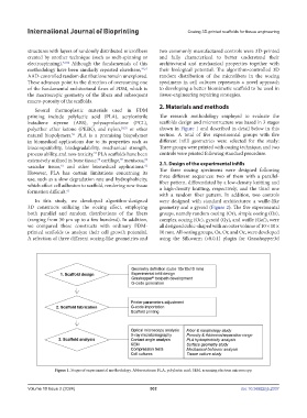
printing include polylactic acid (PLA), acrylonitrile The research methodology employed to evaluate the
butadiene styrene (ABS), polycaprolactone (PCL), scaffolds design and microstructure was based in 3 stages
polyether ether ketone (PEEK), and nylon, 20,55 or other shown in Figure 1 and described in detail below in this
56
natural biopolymers. PLA is a promising biopolymer section. A total of five experimental groups with five
in biomedical applications due to its properties such as different infill geometries were selected for the study:
biocompatibility, biodegradability, mechanical strength, Three groups were printed with oozing technique, and two
process ability, and non-toxicity. PLA scaffolds have been controls were printed following standard procedure.
57
extensively utilized in bone tissue, cartilage, meniscus, 2.1. Design of the experimental infills
58
59
60
vascular tissue, and other biomedical applications.
61
62
However, PLA has certain limitations concerning its The three oozing specimens were designed following
use, such as a slow degradation rate and hydrophobicity, three different sequences: two of them with a parallel-
which affect cell adhesion to scaffold, rendering new tissue fiber pattern, differentiated by a low-density knitting and
formation difficult. 61 a high-density knitting, respectively, and the third one
with a random fiber pattern. In addition, two controls
In this study, we developed algorithm-designed were designed with standard architectures: a waffle-like
3D constructs utilizing the oozing effect, employing geometry and a gyroid (Figure 2). The five experimental
both parallel and random distributions of the fibers groups, namely random oozing (Or), simple oozing (Os),
(ranging from 30 µm up to a few hundred). In addition, complex oozing (Oc), gyroid (Gy), and waffle (Gof), were
we compared these constructs with ordinary FDM- all designed cubic-shaped with an outer volume of 10 × 10 ×
printed scaffolds to analyze their cell growth potential. 10 mm. All oozing groups, Os, Or, and Oc, were developed
A selection of three different oozing-like geometries and using the Silkworm (v0.0.1) plugin for Grasshopper3d
Figure 1. Stages of experimental methodology. Abbreviations: PLA, polylactic acid; SEM, scanning electron microscopy.
Volume 10 Issue 2 (2024) 502 doi: 10.36922/ijb.2337