
Page 502 - IJB-10-2
P. 502
International Journal of Bioprinting Mineralization of 3D-printed PHA scaffolds
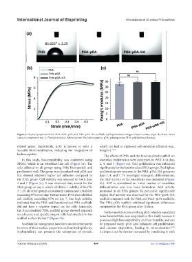
Figure 3. Physical properties of the PHA, PHA–pDA, and PHA–pDA–HA scaffolds. (a) Representative images of water contact angle. (b) Stress–strain
curve of compression test. (c) Young’s modulus. Abbreviations: HA, hydroxyapatite; pDA, polydopamine; PHA, polyhydroxyalkanoate.
related genes. Importantly, pDA is known to offer a which can lead to improved cell–substrate adhesion (e.g.,
versatile biomineralization, including the integration of integrin). 17,18
hydroxyapatite.
The effects of PHA and the functionalized scaffold on
In this study, biocompatibility was confirmed using osteoblast proliferation were confirmed by WST-1 on days
MG63, which is an osteoblast-like cell (Figure 5a). The 1, 4, and 7 (Figure 6a). Cell proliferation was enhanced
cells adhered to all groups using PHA biomaterials and significantly for the functionalized PHA groups. The highest
proliferated well. The group functionalized with pDA and proliferation rate was seen in the PHA–pDA–HA group on
HA showed relatively higher cell adhesion compared to days 1, 4, and 7. To investigate osteogenic differentiation,
the PHA group. Cell viability was assessed on both days the ALP activity of the osteoblasts was measured (Figure
4 and 7 (Figure 5c). It was observed that, except for the 6b). ALP is considered an initial marker of osteoblast
PHA group on day 4, which exhibited a viability of 86.67% differentiation and new bone formation. ALP activity
± 1.29, all other groups consistently maintained a viability increased in all PHA groups. In particular, significantly
exceeding 97% every day. Furthermore, PHA also exhibited higher ALP activity was observed for the PHA–pDA–HA
cell viability exceeding 97% on day 7. The high viability scaffold compared with the PHA and PHA–pDA scaffolds.
indicated that the PHA and functionalized PHA scaffolds The PHA–pDA scaffold exhibited significant differences
did not have a negative impact on the cells. Especially, compared to the PHA group after 10 days.
the functionalized PHA scaffold group showed apparent Surface modification involving pDA, which is a qualified
enrichment, and spindle-shaped cells had attached to the bone biosubstitute, was employed in this study because it
scaffold surface by day 7 (Figure 5b). possesses high biocompatibility and functionality, and can
Scaffolds for osteogenesis have been studied extensively be prepared easily. pDA also enhances cell proliferation
in terms of their surface properties, such as hydrophilicity. and calcium deposition, leading to mineralization. 32,34
Hydrophilicity can promote the adsorption of protein, Its impact can be further increased by combining it with
Volume 10 Issue 2 (2024) 494 doi: 10.36922/ijb.1806