
Page 504 - IJB-10-2
P. 504
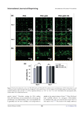
International Journal of Bioprinting                               Mineralization of 3D-printed PHA scaffolds


            Figure 5. Viability of osteoblast-like cells on the PHA, PHA–pDA, and PHA–pDA–HA scaffolds. (a) Live and dead assays of MG63 cells on days 1, 4, and
            7. (b) Representative magnified images depicting cell adhesion on the scaffold on day 7. (c) Cell viability of the polyhydroxyalkanoate (PHA) and surface-
            modified scaffolds on day 4 and day 7. n = 4; NS, no significant difference; *p < 0.05; **p < 0.01; ***p < 0.001. Abbreviations: HA, hydroxyapatite; pDA,
            polydopamine; PHA, polyhydroxyalkanoate.

            growth factors.  Therefore, coating the PHA surface   similar to the mineral phase of bone. 16,36  Thus, HA-based
                         35
            with pDA facilitates osteoconductivity by enhancing cell   bone  scaffolds  enhance  the  osteogenic  differentiation
            proliferation and differentiation. Also, biocompatible HA   of osteoblast-like cells and promote bone repair both  in
            is generally used for bone scaffolds, as its composition is   vitro and in vivo. 33,37-39  The results of this study confirmed


            Volume 10 Issue 2 (2024)                       496                                doi: 10.36922/ijb.1806