
Page 73 - IJB-3-1
P. 73
Swee Leong Sing, Shuai Wang, Shweta Agarwala, et al.
(A) (B)
(C) (D)
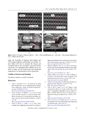
Figure 3. Micro-CT images of biphasic scaffolds (A) cpTi (B) TiTa, and SEM images of (C) cpTi and (D) TiTa, showing infiltration of
Type 1 collagen into the pores
study, the feasibility of forming cpTi-collagen and tilage and subchondral bone regeneration. Knee Surgery
TiTa-collagen biphasic scaffolds has been shown. Fu- Sports Traumatology Arthroscopy, vol.20(6): 1182±1191.
ture studies will aim to optimize the designs and https://doi.org/10.1007/s00167-011-1655-1
evaluation with in vitro cell culture experiment will be 4. Wang S, Taraballi F, Tan L P, et al., 2012, Human kera-
carried out. It is anticipated that scaffolds can be tai- tin hydrogels support fibroblast attachment and prolifer-
lored to better suit the biochemical and mechanical ation in vitro. Cell and Tissue Research, vol.347(3): 795±
requirements for osteochondral tissue regeneration. 802.
https://doi.org/10.1007/s00441-011-1295-2
Conflict of Interest and Funding 5. Wang S, Wang Z, Foo S E M, et al., 2015, Culturing Fi-
broblasts in 3D Human Hair Keratin Hydrogels. ACS
The authors declare no conflict of interest.
Applied Materials & Interfaces, vol.7(9): 5187±5198.
References https://doi.org/10.1021/acsami.5b00854
6. Hollister S J, 2005, Porous scaffold design for tissue en-
1. Csaki C, Schneider P R A , and Shakibaei M, 2008, gineering. Nature Materials, vol.4(7): 518±524.
Mesenchymal stem cells as a potential pool for cartilage https://doi.org/10.1038/nmat1421
tissue engineering. Annals of Anatomy-Anatomischer 7. Duan X, Zhu X, Dong Z, et al., 2013, Repair of large
Anzeiger, vol.190(5): 395±412. osteochondral defects in a beagle model with a novel
https://doi.org/10.1016/j.aanat.2008.07.007 type I c ollagen/glycosaminoglycan-porous titanium bi-
2. Kon E, Filardo G, Di Martino A, et al., 2014, Clinical phasic scaffold. Materials Science & Engineering C-
results and MRI evolution of a nano-composite multi- Materials for Biological Applications, vol.33(7): 3951±
layered biomaterial for osteochondral regeneration at 5 3957.
years. American Journal of Sports Medicine, vol.42(1): https://doi.org/10.1016/j.msec.2013.05.040
158±165. 8. Nover A B, Lee S L, Georgescu M S, et al., 2015, Po-
https://doi.org/10.1177/0363546513505434 rous titanium bases for osteochondral tissue engineering.
3. Panseri S, Russo A, Cunha C, et al., 2012, Osteo- Acta Biomaterialia, vol.27: 286±293.
chondral tissue engineering approaches for articular car- https://doi.org/10.1016/j.actbio.2015.08.045
International Journal of Bioprinting (2017)–Volume 3, Issue 1 69