
Page 71 - IJB-3-1
P. 71
Swee Leong Sing, Shuai Wang, Shweta Agarwala, et al.
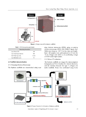
Figure 1. Design concept for biphasic scaffolds
Table 1. SLM processing parameters ning electron microscopy (SEM), using a s canning
Processing Parameters electron microscope (JEOL JSM-5600LV, Japan). Sca-
ffolds were frozen at −20 °C for two days and lyophi-
Laser Power (W) 100
Laser Scan Speed (mm/s) 500 lized. All samples were gold-sputtered at 18 mA for
10 sec. Images were taken at an accelerating voltage
Hatch Spacing (mm) 0.120
of 10 kV under high vacuum.
Layer thickness (µm) 30
2.3.2 Micro-CT evaluation
2.3 Scaffold characterization The biphasic scaffolds are imaged by microcomputed
tomography (µCT) to visualize the internal structures,
2.3.1 Scanning Electron Microscopy
and the interface between the type 1 collagen and
The biphasic scaffolds are characterized using scan- metal scaffolds. Scans were performed using X-ray
Figure 2. Process flowchart for fabrication of biphasic scaffolds
International Journal of Bioprinting (2017)–Volume 3, Issue 1 67