
Page 304 - IJB-10-3
P. 304
International Journal of Bioprinting Design and optimization of 3DP bioscaffolds
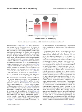
Figure 13. Cell counts for the multi-channel scaffolds with different volume fraction of channel on day 7.
fraction equaled to 7.5% (Figure 12e). This is attributed to exhibited the highest cell numbers on day 7 compared to
the interplay between the increase in cell density and the others, confirming the effectiveness of this optimization
reduction of scaffold materials for carrying cells. Note that method.
this value may vary when other parameters differ, such as Channels or voids are key architectural features in
the height and diameter of the scaffold, cell type, scaffold scaffolds, providing a passageway to guide cell growth
material, and nutrient flow conditions. The second step is and facilitating nutrient and waste exchange. 11,14,15 The
to spread the single-channel volume fraction (7.5% for this degree of cellularity in scaffolds is critical to ensure the
case) to the scaffold to form a multi-channeled scaffold biological functionalities of the engineering tissue, and
with optimal geometric parameters, including channel highly cellularized scaffolds can enhance intercellular and
diameter and number (see Figure 12f). When the number cell–material interactions, therefore improving cellular
of channels increased from 1 to 21, the normalized average behaviors. During the design of porous scaffolds, adding
45
oxygen concentration rose from 23.2% to 40.95% (Figure more empty phases can improve the permeability and thus
12g). The volume fraction of the hypoxic region, where the cell proliferation; however, it reduces the solid volume for
oxygen concentration was lower than the threshold C <14 carrying cells. The correct geometric parameters of scaffolds
μM, decreased from 46.02% to only 8.18% (Figure 12h). yielding maximum cell number should be determined, in
20
As a result, the number of cells elevated from 1,082,166 to order to maximize the cellularity degree of the scaffolds.
1,492,918, showing an increment of 37.96% (Figure 12i However, the selection of channel parameters is somewhat
and j). The specific surface area is a critical indicator to arbitrary in current practice or based on experiments, and
characterize the performance of scaffold as it influences the current design rules are insufficient to obtain the most
the rate of oxygen and nutrients exchange significantly. optimal parameters. The results showed that the channel
The specific surface area increases when increasing the number and size significantly impact cell proliferation.
void volume fraction (Figure 12a) or splitting the empty Simply adding channels improves oxygen transportation
phase into more tiny channels (Figure 12f). The specific and cell density. However, excessive channels will reduce
surface area is enlarged from 1.5 to 2.35 when the number the scaffold materials and thus the final cell number. The
of channels increases from 1 to 21 (Figure 12f), indicating modeling-based optimization strategy proposed in this
that scaffolds featuring more tiny channels possess better study can determine the optimal channel parameters that
permeability and therefore higher cell growth rates. This ensure maximum cell number after culturing. For this
is consistent with the modeled cell density (Figure 12i), case, the optimal scaffold features 21 uniformly distributed
further verifying the correctness of the model. To validate channels with a diameter of 0.3 mm. Theoretically, the
that the structure with multiple channels was optimal, a cell count can be further improved by increasing the
similar procedure was applied for scaffolds with channel channel number to more than 21. Scaffolds with desired
volume fractions of 5% and 10% for comparison. As shown permeability and cell proliferation are usually geometrically
in Figure 13, the scaffolds with a volume fraction of 7.5% complex and feature microsized channels and thin walls,
Volume 10 Issue 3 (2024) 296 doi: 10.36922/ijb.1838