
Page 574 - IJB-10-3
P. 574
International Journal of Bioprinting Bioprint micro breast cancer
Figure 5. Microvasculature development within printed micro-cancer tissues (PMCaTs). (A) Architecture of the micro-cancer tissue, highlighting breast
cancer cells (green), intricate microvasculature (red), and fibroblasts (blue). (B and C) Depiction of cancer cells (green) and microvasculature channels
(red). Scale bars: 100 μm.
Such an observation not only deepens our used AlexaFluo488-Dextran with a molecular weight
understanding of tumor biology but also paves the way of 70 kDa as a surrogate. This molecular weight is
for novel therapeutic avenues. Given the central role of approximately half that of many therapeutic antibodies,
the microvascular system in tumor progression, targeting suggesting its diffusion could be even slower than actual
it could prove to be groundbreaking. Future therapies antibodies. Our experiments provide intriguing insights
may not only aim to inhibit cancer cell growth but also into its penetration within PMCaTs. As demonstrated
target the cancer microvasculature, potentially arresting in Figure 6A–C, the dye effectively penetrated the
metastatic progression and offering a more comprehensive cell over time, suggesting time-dependent diffusion
approach to cancer treatment. 26,27 characteristics of the modeled drug.
3.6. Dynamics of drug penetration Interestingly, the rate and pattern of dye diffusion
The penetration dynamics of therapeutics, especially in the PMCaTs provide cues to the matrix’s inherent
those of high molecular weight, remain an area of characteristics. One of the significant distinguishing
intense research, particularly in the context of solid features of PMCaTs, as compared to other models that
tumors. Many current therapies, such as monoclonal embed cancer cells in hydrogel or rely solely on pure cell
antibodies in immunotherapy or targeted therapy, have aggregates, is the presence of a considerable amount of
molecular weights around 150 kDa, while traditional relatively rigid matrix. Such a matrix can act as a barrier,
chemotherapy agents are much smaller, typically potentially modulating the diffusion dynamics of drugs,
ranging from 200 to 900 Da. Given the importance of slowing down their penetration, and influencing the
understanding the penetration of larger molecules, we overall therapeutic efficacy.
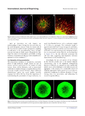
Figure 6. Modeling drug penetration using AlexaFluo488-Dextran (70 kDa). Evaluation of the depth of penetration of the fluorescent dye AlexaFluo488-
Dextran, employed here as a representative model for large molecular weight compounds such as antibodies. Scale bars: 100 μm.
Volume 10 Issue 3 (2024) 566 doi: 10.36922/ijb.2911