
Page 575 - IJB-10-3
P. 575
International Journal of Bioprinting Bioprint micro breast cancer
This unique matrix structure, while making PMCaTs representative platform for studying these challenges.
more physiologically relevant by mimicking the natural The interactions between T cells and cancerous tissues
tumor environment, also poses challenges for drug in our model provide insights into their engagement
delivery. The intricate interplay between the rigidity and potential neutralization mechanisms. However,
of the matrix and the diffusion of large molecules may while the model presents a valuable foundation, it is
elucidate some of the obstacles encountered in clinical in its initial stages. Given the growing importance of
settings, especially when administering antibody-based immunotherapy, our findings highlight the need for
therapies. Moreover, this has broader implications. The rigorous validation, incorporation of varied immune
understanding that PMCaTs can accurately simulate drug cell dynamics, and evaluations in drug testing scenarios
penetration due to their matrix composition emphasizes to ensure the model’s relevance and applicability in
the need for refined drug delivery techniques tailored for therapeutic studies.
specific tumor microenvironments.
3.8. Modeling cancer metastasis
Going forward, these findings stress the importance of Metastasis, the spread of cancer cells from the primary
not only targeting cancer cells but also understanding and tumor to distant sites, is a key factor in cancer progression
possibly manipulating the tumor microenvironment for and the leading cause of cancer-related deaths. 33,34 By
better therapeutic outcomes. It might be worth exploring focusing on the dynamics of metastasis, our study aims to
strategies to modulate matrix rigidity or enhance drug shed light on the mechanisms underlying cancer spread,
penetration by changing the tumor matrix architecture, offering potential avenues for therapeutic intervention.
28
offering a two-pronged approach to tackle tumor resistance
and ensure a more effective drug delivery. 29 Utilizing PMCaTs as our experimental model, we
sought to visualize and understand the dynamics of
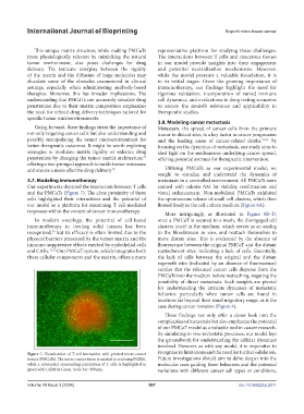
3.7. Modeling immunotherapy metastasis in a controlled environment. All PMCaTs were
Our experiments depicted the interaction between T cells stained with calcein AM for viability confirmation and
and the PMCaTs (Figure 7). The close proximity of these visual enhancement. Non-mobilized PMCaTs exhibited
cells highlighted their interactions and the potential of the spontaneous release of small cell clusters, which then
our model as a platform for examining T cell-mediated floated freely in the cell culture medium (Figure 8A).
responses within the context of cancer immunotherapy.
More intriguingly, as illustrated in Figure 8B–D,
In modern oncology, the potential of cell-based once a PMCaT is secured to a mesh, the disengaged cell
immunotherapy in treating solid tumors has been clusters travel in the medium, which serves as an analog
recognized, but its efficacy is often limited due to the to the bloodstream in vivo, and reattach themselves to
30
physical barriers presented by the tumor matrix and the more distant sites. This is evidenced by the absence of
immune-suppressive effects exerted by endothelial cells fluorescence between the original PMCaT and the distant
and CAFs. 31,32 Our PMCaT system, which integrates both reattachment sites, indicating a lack of cells. Essentially,
these cellular components and the matrix, offers a more the lack of cells between the original and the distant
regrowth sites (indicated by an absence of fluorescence)
verifies that the relocated cancer cells disperse from the
PMCaTs into the medium before reattaching, negating the
possibility of direct metastasis. Such insights are pivotal
for understanding the intricate dynamics of metastatic
behavior, particularly when tumor cells are found in
locations far beyond their usual migratory range, as is the
case during cancer invasion (Figure 4).
These findings not only offer a closer look into the
complexities of metastasis but also emphasize the potential
of our PMCaT model as a valuable tool in cancer research.
By simulating in vivo metastatic processes, our model lays
the groundwork for understanding the cellular dynamics
involved. However, as with any model, it is imperative to
Figure 7. Visualization of T cell interaction with printed micro-cancer recognize its limitations and the need for further validation.
tissues (PMCaTs). The micro-cancer tissue is marked in red using PKH26, Future investigations should aim to delve deeper into the
while a substantial surrounding population of T cells is highlighted in molecular cues guiding these behaviors and the potential
green with CellBrite Green. Scale bar: 300 μm. variations with different cancer cell types or conditions,
Volume 10 Issue 3 (2024) 567 doi: 10.36922/ijb.2911