
Page 576 - IJB-10-3
P. 576
International Journal of Bioprinting Bioprint micro breast cancer
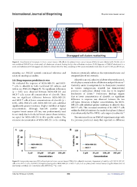
Figure 8. Visualization of metastasis in micro-cancer tissues. All cells in the printed micro-cancer tissues (PMCaTs) were labeled with PKH26. (A) A
non-mobilized PMCaT from which small cell clusters are released, floating freely in the cell culture medium. (B–D) Sequence of PMCaT attachment to a
mesh and reattachment of disengaged cell clusters to distant sites over time, emulating in vivo cancer metastasis. Scale bars: (A and C) 100 μm; (D) 50 μm.
ensuring our PMCaT system’s continued relevance and literature potentially enhances the representativeness and
utility in oncological studies. comparability of our research.
3.9. Drug response prediction in vivo Alisertib is an oral, selective inhibitor of aurora kinase A,
35
We evaluated the response of MDA-MB-231 and MCF- which plays a crucial role in cell division and proliferation.
7 cells to alisertib in both traditional 2D cultures and Currently under investigation for its therapeutic potential
within our PMCaTs (Figure 9). No significant differences in various malignancies, alisertib has demonstrated
(p > 0.05) were observed between MDA-MB-231 and promise in early-phase clinical trials due to its targeted
36
MCF-7 cells across all concentrations of alisertib. There mechanism of action. Preliminary findings suggest
was no significant difference between MDA-MB-231 that at lower concentrations of alisertib, no significant
and MCF-7 cells at lower concentrations of alisertib (p > difference in response was observed between the two
0.05), while PMCaTs with MDA-MB-231 cells exhibited cell types. However, at higher concentrations, the MDA-
significantly greater resistance (higher viability) at higher MB-231 cells exhibited greater resistance to alisertib than
concentrations. Although Sum149, another typical MCF-7 cells. This increased sensitivity of the MCF-7 cells
negative breast cancer cell line, was predominantly used within the PMCaTs mirrors the results from phase 2 clinical
37
in our study due to its ductal breast cancer characteristics, trials where only the ER+/HER2- cells respond to alisertib.
we opted for MDA-MB-231 in this specific section. The The outcomes from our PMCaT experiments align with
extensive documentation of MDA-MB-231 in the existing the previous preclinical study that reported no difference
Figure 9. Comparative response of 2D cells and printed micro-cancer tissues (PMCaTs) to alisertib treatment. Assessment of viability in 2D MDA-MB-231
cells versus MCF-7 cells, and PMCaTs bioprinted using either MDA-MB-231 or MCF-7 cells, both co-printed with normal human lung fibroblasts (NHLF)
and human umbilical vein endothelial cells (HUVECs). ***p < 0.05.
Volume 10 Issue 3 (2024) 568 doi: 10.36922/ijb.2911