
Page 58 - IJB-10-3
P. 58
International Journal of Bioprinting 3D-printed biodegradable metals for bone regeneration
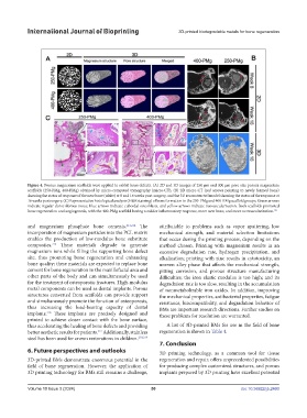
Figure 4. Porous magnesium scaffolds were applied to rabbit bone defects. (A) 2D and 3D images of 250 μm and 400 μm pore size porous magnesium
scaffolds (250-PMg, 400-PMg) obtained by micro-computed tomography (micro-CT), (B) 2D micro-CT (red arrows pointing to newly formed bone)
showing the status of response of the new bone (white) at 8 and 16 weeks post-surgery, and the 3D reconstructed model showing the status of the response at
16 weeks postsurgery. (C) Representative histological analysis (H&E staining) of bone formation in the 250-PMg and 400-PMg scaffold groups. Green arrows
indicate regular dense fibrous tissue, blue arrows indicate cuboidal osteoblasts, and yellow arrows indicate neovascularization. Both scaffolds promoted
bone regeneration and angiogenesis, with the 400-PMg scaffold having a milder inflammatory response, more new bone, and more neovascularization. 125
and magnesium phosphate bone cements. 213,214 The attributable to problems such as vapor sputtering, low
incorporation of magnesium particles into the PCL matrix mechanical strength, and material selection limitations
enables the production of low-modulus bone substitute that occur during the printing process, depending on the
215
composites. These materials degrade to generate method chosen. Printing with magnesium results in an
magnesium ions while filling the supporting bone defect excessive degradation rate, hydrogen precipitation, and
site, thus promoting bone regeneration and enhancing alkalization; printing with zinc results in cytotoxicity, an
bone quality; these materials are expected to replace bone uneven alloy phase that affects the mechanical strength,
cement for bone regeneration in the maxillofacial area and pitting corrosion, and porous structure manufacturing
other parts of the body and can simultaneously be used difficulties; the iron elastic modulus is too high; and its
for the treatment of osteoporotic fractures. High-modulus degradation rate is too slow, resulting in the accumulation
metal components can be used as dental implants. Porous of nonmetabolizable iron oxides. In addition, improving
structures converted from scaffolds can provide support the mechanical properties, antibacterial properties, fatigue
and simultaneously promote the function of osteoporosis, resistance, biocompatibility, and degradation behavior of
thus increasing the load-bearing capacity of dental BMs are important research directions. Further studies on
implants. These implants are precisely designed and these problems for resolution are warranted.
216
printed to achieve closer contact with the bone surface,
thus accelerating the healing of bone defects and providing A list of 3D-printed BMs for use in the field of bone
better aesthetic results for patients. Additionally, stainless regeneration is shown in Table 4.
217
steel has been used for crown restorations in children. 218,219
7. Conclusion
6. Future perspectives and outlooks 3D printing technology, as a common tool for tissue
3D-printed BMs demonstrate enormous potential in the regeneration and repair, offers unprecedented possibilities
field of bone regeneration. However, the application of for producing complex customized structures, and porous
3D printing technology for BMs still remains a challenge, implants prepared by 3D printing have excellent potential
Volume 10 Issue 3 (2024) 50 doi: 10.36922/ijb.2460