
Page 59 - IJB-10-3
P. 59
International Journal of Bioprinting 3D-printed biodegradable metals for bone regeneration
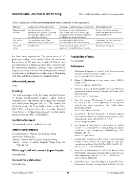
Table 4. Applications of 3D-printed biodegradable metals in the field of bone regeneration
Materials Mechanisms for bone regeneration Advantages and disadvantages in application Medical applications
Magnesium and Secreting osteogenic cytokines, Density and Young’s modulus close to Fracture, bone defect, cartilage
its alloys synergizing with cytokines, promoting those of bone tissue, but excessively high repair, bone substitution composite
osteogenic gene expression, and biodegradation rate, hydrogen precipitation material additives, etc.
activating related pathways and alkalization during degradation
Zinc and its Zinc homeostasis, redox effect, Good antibacterial effect, but low Osteoporotic fractures, bone defect
alloys antibacterial ability, vascularization biocompatibility and difficult processing repair and dentistry, etc.
Iron and its Vascularization Strong mechanical support, but low Nanoparticle coating, etc.
alloys degradation rate and high elastic modulus
for bone tissue regeneration. The advancement of 3D Availability of data
printing technology and materials leads to the continuous
improvement of BM materials. 3D-printed BMs are used Not applicable.
not only for fracture fixation and bone defect repair but also References
for osteoporotic fractures, cartilage repair, maxillofacial
surgery, and other processes. We anticipate more research 1. Wildemann B, Ignatius A, Leung F, et al. Non-union bone
contributions regarding further applications of 3D printing fractures. Nat Rev Dis Primers. 2021;7(1):57.
with BMs and their expansion to unexplored fields. doi: 10.1038/s41572-021-00289-8
Acknowledgments 2. Megas P. Classification of non-union. Injury. 2005;36
(Suppl 4):S30-37.
None. doi: 10.1016/j.injury.2005.10.008
3. Dimitriou R, Jones E, McGonagle D, Giannoudis PV. Bone
Funding regeneration: current concepts and future directions. BMC
This work was supported by the Young Scientist Program Med. 2011;9:66.
doi: 10.1186/1741-7015-9-66
of Beijing Stomatological Hospital, Capital Medical
University (No. YSP202208); the National Key Research 4. Daly AC, Freeman FE, Gonzalez-Fernandez T, Critchley
and Development Program (No. 2022YFA1104401); the SE, Nulty J, Kelly DJ. 3D bioprinting for cartilage and
CAMS Innovation Fund for Medical Sciences (No. 2019- osteochondral tissue engineering. Adv Healthc Mater.
2017;6(22):1700298.
I2M-5-031); and grants from the Innovation Research doi: 10.1002/adhm.201700298
Team Project of Beijing Stomatological Hospital, Capital
Medical University (No. CXTD202204). 5. Liao J, Wu S, Li K, Fan Y, Dunne N, Li X. Peptide-modified
bone repair materials: factors influencing osteogenic activity.
Conflict of interest J Biomed Mater Res A. 2019;107(7):1491-1512.
doi: 10.1002/jbm.a.36663
The authors declare no conflicts of interest. 6. Sanz-Sanchez I, Sanz-Martin I, Ortiz-Vigon A, Molina
A, Sanz M. Complications in bone-grafting procedures:
Author contributions classification and management. Periodontology 2000.
Conceptualization: Zhipeng Fan, Lingxiao Wang 2022;88(1):86-102.
Supervision: Zhipeng Fan doi: 10.1111/prd.12413
Writing – original draft: Yang Liu, Lingxiao Wang 7. Graham SM, Leonidou A, Aslam-Pervez N, et al. Biological
Writing – review & editing: Lingxiao Wang, Yang Liu, therapy of bone defects: the immunology of bone allo-
Zhipeng Fan transplantation. Expert Opin Biol Ther. 2010;10(6):885-901.
doi: 10.1517/14712598.2010.481669
Ethics approval and consent to participate 8. Bavya Devi K, Lalzawmliana V, Saidivya M, Kumar V, Roy
M, Nandi SK. Magnesium phosphate bioceramics for bone
Not applicable. tissue engineering. Chem Rec. 2022;22(11):e202200136.
doi: 10.1002/tcr.202200136
Consent for publication
9. Sordi MB, Cruz A, Fredel MC, Magini R, Sharpe PT. Three-
Not applicable. dimensional bioactive hydrogel-based scaffolds for bone
Volume 10 Issue 3 (2024) 51 doi: 10.36922/ijb.2460