
Page 135 - IJB-4-2
P. 135
Fabrication of biomimetic placental barrier structures within a microfluidic device utilizing two-photon polymerization
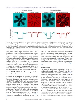
Figure 6. Color gradient of riboflavin and dextran shows permeability of the membrane with different wall thicknesses.The color intensity
was depicted along a linear axis (white). Thereby the color intensity (AU) of pixels on this line was measured and plotted using the
ZEN software. Thereby two different wall thicknesses were analyzed. 25 µm on the left side and 100 µm on the right side. The potential
to retain high molecular weight substances was demonstrated using dextran (blue) as in both samples the inner fluorescence signal is
approaching zero. Riboflavin (red) on the other side is a membrane permeable substance, shown by equal color intensity within and
outside the villous structure.
sides, while dextran stayed exclusively outside of the GelMOD-AEMA membrane, where cells adhered to the
membrane. Direct comparison of signal differences channel wall as well as to the chip material. The green
between inner and outer region numerical values, tint of the membrane is a result of the high fluorescence
indicate a marginally improved diffusion capacity of the structured material. The cell layer around the
of riboflavin in samples with smaller wall thickness. membranous structure is even denser 7 days after
25 µm samples can be recognized by a significant seeding, which can be seen in Figure 7B. When enlarged
smaller transition zone compared to 100 µm villi- it becomes clear that imaged cells are in different layers,
samples. Here again the dextran signal was detected as some of them are more blurred than others (Figure
exclusively outside of the structure while riboflavin 7C).
levels can be considered equal on both sides. In the 4. Discussion
range of thickness and 2PP parameters used in this study,
no substantial dependence of diffusion was observed. The necessity of reliable in vitro models in the field
3.6 GelMOD-AEMA Membrane Supports Cell of placental research is well reflected by the fact that
[17–19,20,21]
.
contradictory data can be found in literature
layer Formation Underlying processes are difficult to study since this
After the establishment of optimal structuring transient organ undergoes constant changes, as it controls
parameters, material composition and cultivation its own growth and functionality at the same time [3,17] .
conditions, membranes were printed followed by seeding Constantly changing structural arrangement, size, and
with HUVECs and BeWo B30 cells successively. Seeded surface area pose high demands on the model and
chips were connected to the microfluidic pump after 24 require a large degree of flexibility. For the simulation
hours of cell settling, to gain preliminary results about of placental processes in vitro, not only the material
the biocompatibility of the model. For cultivation, the biocompatibility but also high adaptability play a role, as
respective channel was supplied with cell type specific the placental geometry changes during pregnancy.
media. After 7 days of cultivation under constant flow, The usage of microfluidic devices in combination
the cell layer formation was determined. Therefore, with 2PP techniques offers a complete new range of
cells were stained with Calcein-AM and imaged using possibilities. Microfluidic setups mimicking entire organs
a confocal microscope. The image depicted in Figure or organisms are gaining more and more acceptance in
7A shows a three-dimensional view of one loop of the the field of tissue engineering and especially in placenta
8 International Journal of Bioprinting (2018)–Volume 4, Issue 2