
Page 215 - IJB-10-4
P. 215
International Journal of Bioprinting 3D-Printed scaffolds for diabetic bone defects
and 3, and the absorbance value at 450 nm was measured min, treated with 0.1% Triton X-100 (Boster, China) for
with an enzyme labeler (Thermo Fisher Scientific Co., Ltd. 15 min, and incubated with rhodamine phalloidin staining
USA). The experiment was repeated three times. solution (50 μg/mL, Boster, China) for 30 min at room
temperature protected from light and then with DAPI
Cell viability was assessed using live/dead cell staining, (Boster, China) for 15 min, and cell adhesion was observed
in which live cells show green fluorescence and dead cells under a laser confocal microscope (Zeiss Axio Observer,
show red fluorescence. BMSCs at passage 4 were transferred Zeiss, Germany). The remaining samples were fixed with
into 24-well plates and co-cultured with different multiwell 2.5% glutaraldehyde (Boster, China) for 3 h. The samples
scaffolds for 48 h. Then, 1 mL of live/dead staining solution were dehydrated in 40%, 50%, 60%, 70%, 80%, 90%, 95%,
(Beyotime, China) was added to each well, and the samples and 100% ethanol solutions for 15 min in sequence before
were incubated at 37°C and 5% CO for 30 min, away from being vacuum-dried. Cell adhesion on the surface of the
2
light, and live and dead cells were observed by fluorescence porous scaffolds was observed by SEM to evaluate the
imaging microscopy. adhesion of composite scaffold materials to BMSCs.
2.3.3. Adhesion and migration of BMSCs to A scratching experiment was conducted to verify
porous scaffolds the effects of different porous scaffolds on the migration
The four porous scaffolds were placed in 24-well plates, and of cells; the preparation of the material extract and the
the BMSCs were inoculated at 2 × 10 /well; then, complete experimental method are illustrated in Figure 3F. First,
4
23
medium was added, and the plates were incubated for BMSCs were inoculated at a density of 2 × 10 /well and
4
48 h at 5% CO and 37°C. A portion of the samples were cultured in 5% CO at 37℃ for 48 h; then, the bottom
2
2
fixed using 4% paraformaldehyde (Boster, China) for 30 surface of the six-well plate was scratched vertically with
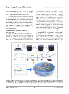
Figure 1. General overview of the study. (A) 3D printing of PCL porous scaffolds, peptide branching, establishment of a PCL@SS31@E7 system with
the capacity to recruit stem cells and regulate mitochondrial respiration, and animal experiments on repair of femoral condylar bone defects in diabetic
rats. (B) PCL porous scaffolds recruit BMSCs using the E7 peptide, regulate mitochondrial respiration function in BMSCs through the SS31 peptide, and
promote the normalization of BMSC function to achieve the principle of bone regeneration.
Volume 10 Issue 4 (2024) 207 doi: 10.36922/ijb.2379