
Page 225 - IJB-10-4
P. 225
International Journal of Bioprinting 3D-Printed scaffolds for diabetic bone defects
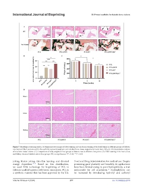
Figure 7. Histological staining results. (A) Representative images of H&E staining and von Kossa staining of the distal femur in different groups of diabetic
rats (red and black portions within the scaffolds represent neoplastic and calcified bone tissue, respectively) (scale bars: 100 μm). (B) Quantitative analysis
of new bone tissue volume. (C) Comparison of body weights of four groups of diabetic rats at different time points. (D) H&E staining of the heart, liver,
and kidney tissues of diabetic rats in four groups. NS, no significance; *P < 0.05; ** P < 0.01.
jetting, binder jetting, thin-film layering, and directed Food and Drug Administration for medical use. Despite
energy deposition. 34-36 Based on this classification, possessing good plasticity and biosafety, its applications
we used FDM technology for bioprinting of PCL to have been limited owing to poor hydrophilicity, a trait
37
fabricate scaffold systems with bionic micropores. PCL is unfavorable for cell attachment. Hydrophilicity can
a synthetic material that has been approved by the U.S. be increased by introducing hydroxyl and carboxyl
Volume 10 Issue 4 (2024) 217 doi: 10.36922/ijb.2379