
Page 237 - IJB-10-4
P. 237
International Journal of Bioprinting N-PLN hydrogels for human skin modeling
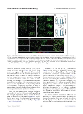
Figure 3. Cell viability and immunofluorescence studies of N-PLN cell-laden hydrogels. (a) Top maximum intensity projections and 3D views at days 1,
3–4, and 7 post-Hs-27 encapsulation for Formulation 1 and Formulation 2 after live/dead assay. Hydrogels were fabricated with these parameters: power =
22 mW, and beam speed = 0.3 mm/s. Live cells stained in green, and dead cells in red. Scale bars = 100 µm. (b) Cell viability quantification based on live/
dead staining at days 1, 3–4, and 7 post-Hs-27 encapsulation for both formulations. Percentages of cell viability are expressed as mean ± SEM (n = 2); ****p
< 0.0001. (c) Immunostaining of Hs-27 within Formulation 2 gels at days 1 (left panel) and 4 (right panel) post-Hs-27 encapsulation: vimentin (green),
collagen IV (red), Ki67 (magenta), nuclei (blue). Scale bars = 200 µm (left) and 100 µm (right).
developed protrusions already since day 1 and looked Expressed at a low level on day 1 (left panel of
spread within the scaffolds (Figure 3a). Overall, these Figure 3c), the staining of collagen IV became more
results seem to point out the superiority of Formulation 2 conspicuous at the cells’ periphery 4 days post-Hs-27
in sustaining the culture of the fibroblasts, probably due to encapsulation, proving the capability of the cells to
the addition of RGD peptides in the polymer composition, produce and secrete the protein (right panel of Figure 3c).
as these are peptide sequences known for promoting Moreover, some nuclei showed positive staining for Ki67,
cell–matrix interactions. Thus, we decided to delve therefore demonstrating the proliferative capacity of the
52
into the functionality of cells growing within hydrogels cells within hydrogels from Formulation 2. Recently, it has
formed from Formulation 2 by immunostaining. To do been reported that N-PLN hydrogels produced by digital
this, we examined proliferative cells by staining for Ki67 light processing technique do not affect cell metabolic
and determining the presence of collagen IV, which is activity nor promote apoptosis. Here, we showed that
53
produced and secreted by the fibroblasts. Cell morphology light-based bioprinting of N-PLN polymers results in
was imaged after vimentin cytoskeleton staining. hydrogels that not only provide good cell viability but
Four days after encapsulation, vimentin was highly also permit cell proliferation and secretion of relevant
expressed in the samples, with the cells totally spread along extracellular matrix proteins.
the hydrogels, especially in the top section (40–50 µm The study on these two formulations underscores the
from the top surface) (right panel in Figure 3c). Indeed, importance to strike a balance between mechanical and
in the upper layers, the cells formed interconnections, biological properties of the gels. Although Formulation
which were not visible yet in the middle layers (Figure S3, 1 could slightly enhance elastic modulus, Formulation 2
Supporting Information). appeared to be a more appropriate choice for supporting
Volume 10 Issue 4 (2024) 229 doi: 10.36922/ijb.3395