
Page 292 - IJB-10-4
P. 292
International Journal of Bioprinting Analysis of PVA-silk fibroin stents
of PVA-SF stents through SC-DW, and discusses the
comprehensive evaluation of the compression properties
and the interaction with the fibroblasts. Finally, the
potential advantages of incorporating SF into PVA for
the development of more effective cardiovascular stents
are highlighted.
2. Methods
2.1. Preparation of polyvinyl alcohol hydrogels
PVA powder (Sigma-Aldrich, United States of America
[USA]), with specifications displayed in Table 1, was mixed
with MilliQ water at a ratio of 28% (w/v). The mixture was
left overnight at 135°C with stirring. It was then stored
at room temperature and preheated to 50°C before its
introduction into the printing syringe. PVA scaffolds
were fabricated using the solvent-cast technique. The PVA
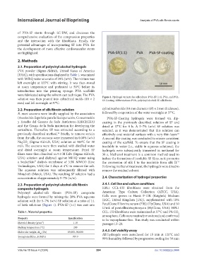
solution was then poured into cylindrical molds (10 × 2 Figure 1. Hydrogel variants for cell culture: PVA-SF (1:1), PVA, and PVA-
SF-Coating. Abbreviations: PVA, polyvinyl alcohol; SF, silk fibroin.
mm) and left overnight at 37°C.
2.2. Preparation of silk fibroin solution cylindrical molds (10 mm diameter (Ø) × 2 mm thickness),
B. mori cocoons were kindly supplied by the association followed by evaporation of the water overnight at 37°C.
(Asociación Española para la Recuperación, Conservación PVA-SF-Coating hydrogels were formed via dip-
y Estudio del Gusano de Seda Autóctono-AERCEGSA) coating in the previously described solution of SF and
and the Granja de la Seda institution for developing the dried at 37°C for 8 h. A 5–7% (w/v) SF solution was
sericulture. Thereafter, SF was extracted according to a selected, as it was demonstrated that this solution can
previously described method. Briefly, to remove sericin effectively coat material surfaces with a very thin layer.
22
23
from the silk, the cocoons were immersed in 0.08% (w/v) A second dip-coating was conducted to ensure consistent
Na CO (Sigma-Aldrich, USA) solution at 100°C for 30 coating of the scaffold. To ensure that the SF coating is
3
2
min. The cocoons were then washed with distilled water insoluble in water (i.e., stable in aqueous solutions), the
and dried overnight at room temperature. Dried SF hydrogels were subsequently immersed in methanol for
fibers were then dissolved in 9.3 M LiBr (Sigma-Aldrich, 30 s. Methanol treatment is a common method used to
USA) solution and dialyzed against MilliQ water using induce the formation of insoluble SF films, as it promotes
a SnakeSkin™ dialysis membrane of 3.5K MWCO (Live the conversion of silk I to the insoluble form silk II.
24
Technologies, USA) for 2 days at 4°C to remove the salt. Following methanol treatment, the hydrogels were dried to
The aqueous solution was subsequently filtered with remove the residual solvent.
Miracloth (Merck, USA). The resulting SF solution had a
concentration of approximately 5–7% (w/w). 2.4. Characterization of hydrogel properties
2.3. Preparation of polyvinyl alcohol-silk fibroin 2.4.1. Cell line and culture conditions
composite hydrogels HFL1 CCL-153 fibroblasts were obtained from the
Polyvinyl alcohol-silk fibroin (PVA-SF) composite American Type Culture Collection (ATCC, USA).
hydrogels were formed by mixing the 28% PVA aqueous Cells were grown in Ham’s F-12K (Kaighn’s) Medium
solution with the 5–7% (w/v) SF solution at a ratio of 1:1 (LGC, United Kingdom [UK]), supplemented with 10%
of both solutions (Figure 1). PVA-SF (1:1) was cast into FetalClone II bovine serum (FBS) (HyClone, USA) and 50
U/mL of penicillin/streptomycin (HyClone, USA). HFL1
Table 1. Material properties CCL-153 fibroblasts were maintained at 37°C and 5% CO
2
atmosphere. Cells were routinely monitored and confirmed
Property Specification to be mycoplasma-free. This study was conducted within
Material density (g/cm³) 1.19 passages 13–20.
Melting temperature (°C ) 200
Molecular weight, M (Da) 89,000–98,000 2.4.2. Cell viability assay
W All hydrogels were autoclaved for 15 min at 121°C and
Young’s modulus, E (MPa) 707.9
90% humidity, followed by progressive cooling for 30 min
Volume 10 Issue 4 (2024) 284 doi: 10.36922/ijb.3444