
Page 293 - IJB-10-4
P. 293
International Journal of Bioprinting Analysis of PVA-silk fibroin stents
to room temperature in a dry environment. Sterilized Table 2. Stent features
scaffolds (10 mm Ø × 2 mm thickness) were placed Feature Specification
in non-adherent cell culture 24-well plates (Sarstedt,
Germany) and soaked in medium supplemented with Geometry Diamond
FBS for 30 min and overnight at 37°C in a 5% CO - Diameter (mm) 5
2
humidified atmosphere prior to cell seeding. This process Strut width (mm) 0.51
facilitates cell attachment and allows us to better assess Hinge width (mm) 1.02
the conditions that would result in higher cell viability. Cell area (mm ) 1.22
2
Approximately, 100,000 cells were suspended in a reduced Number of cells 38
volume of medium (60–100 µL) and were seeded over the
center of the stents using the immersion method. Finally,
seeded scaffolds were incubated for 3 days to allow cell 2.5.1. Compression test
attachment and proliferation. The medium was aspirated A compression resistance parallel plate test was performed
and washed twice with phosphate-buffered saline (PBS) to measure the stents’ radial strength (Figure 3). Stents
(Hyclone, USA). were compressed using a modular compact rheometer
MCR 302e (Anton Paar, Canada) equipped with a load cell
This ensured the removal of crystals not adhering of 50 N, with the upper plate advancing towards the lower
to the surface and ensured that only crystals adhered plate at 0.6 mm/min, following ISO 25539-2 standards
to the stent were counted. Thereafter, 1 mL of medium (n = 6). At a 50% reduction in diameter, the radial force was
26
and 100 µL MTT (Sigma-Aldrich, USA) were added, measured continuously throughout the remaining cycle.
and samples were incubated for 150 min. After
incubation, formazan crystals were dissolved with 1 2.5.2. Colonization assay
mL DMSO (Sigma-Aldrich, USA) with shaking. Four An MTT assay was performed to verify the correct
100 µL aliquots from each well were pipetted into a attachment of cells to the stents and determine the
96-well plate and placed into a microplate reader (Bio- proliferation and colonization of fibroblasts along the
Rad, USA). Absorbance was measured at 570 nm. stent. PVA stents were manufactured and soaked in FBS
Electrospun 15% polycaprolactone (PCL) scaffolds overnight in non-adherent cell culture 12-well plates
were used as a reference and fabricated according to (Sarstedt, Germany) at 37°C and in a 5% CO -humidified
2
previously established protocols. These scaffolds were atmosphere prior to cell seeding. Approximately, 100,000
seeded in non-adherent cell culture plates with the same cells were then prepared in a reduced volume of medium
cell density used for the hydrogel cultures. 25 (60–100 µL) and deposited by the immersion method
2.4.3. Dynamic mechanical analysis of hydrogels
Mettler Toledo DMA/SDTA 861 (Mettler Toledo, USA),
equipped with dual cantilever tools, was used to perform
dynamic mechanical analysis (DMA). The test was run
with a preload of 1 N and a frequency of 1 Hz. The samples
(n = 2) were heated from 25 to 180°C at 5°C/min in an
ambient atmosphere.
2.5. Stent preparation and characterization
PVA stents were printed using a 3D printing technology
(home made DIW 3D printer) based on a rotating
mandrel, as presented in previous studies. Specifically, in
2
this study, the printing head was replaced by a 1-mL Luer-
Lok Tip syringe (BD, USA). The ink was ejected from the
syringe through a mechanical piston that controlled the
volume inside the reservoir. The ink was extruded through
a precision G21 gauge needle (Nordson EFF, USA), which
had an inner diameter of 0.51 mm. The fabrication of
the stents was conducted at 25ºC throughout the entire
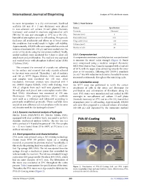
process. Table 2 summarizes the specifications of the stent. Figure 2. Visualization of PVA-SF-Coating (left) and PVA (right)
Thereafter, the PVA stents were dip-coated to fabricate the stents: longitudinal (top) and axial (bottom) views. Abbreviations: PVA,
PVA-SF-Coating stents (Figure 2). polyvinyl alcohol; SF, silk fibroin.
Volume 10 Issue 4 (2024) 285 doi: 10.36922/ijb.3444