
Page 297 - IJB-10-4
P. 297
International Journal of Bioprinting Analysis of PVA-silk fibroin stents
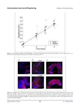
Figure 6. Cell proliferation analysis of HFL1 fibroblasts on PVA-SF-Coating stents and electrospun PCL scaffolds at various time points (2, 5, 7,
and 9 days). The mean and standard deviations are plotted at each time point (n = 3).
Figure 7. Images depicting HFL1 fibroblasts cultured on PVA-SF-Coating stents for 5 (top) and 9 (bottom) days, captured using a confocal laser scanning
microscope. (a) HFL1 fibroblasts observed at a magnification of x20. (b) HFL1 fibroblasts observed at a magnification of x10. Actin cytoskeleton was
stained with rhodamine-phalloidin (red), and nuclei were stained with DAPI (blue). Scale bars: 200 µm (a and b). (c) A 3D reconstruction from a series
of confocal microscope images taken at different Z-stacks to present an overview of the cells on the stent surface. Scale bars: XXX. Abbreviations: PVA,
polyvinyl alcohol; SF, silk fibroin.
Volume 10 Issue 4 (2024) 289 doi: 10.36922/ijb.3444