
Page 295 - IJB-10-4
P. 295
International Journal of Bioprinting Analysis of PVA-silk fibroin stents
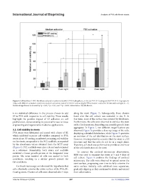
Figure 4. Cell viability of HFL1 fibroblasts cultured on polyvinyl alcohol (PVA) hydrogels, i.e., in the (a) PVA-SF-Coating and (b) PVA-SF (1:1) groups, for
3 days with different activation conditions: medium activation: soaked for 30 min and overnight; FBS activation: soaked for 30 min and overnight (n = 4).
Statistical significance is denoted by *p < 0.05, **p < 0.01, and ***p < 0.001. Abbreviation: SF, silk-fibroin.
is no statistical difference in the process chosen to add along the stent (Figure 7). Subsequently, these clusters
SF to PVA with respect to its cell viability. These results fused after the cell culture was extended to day 9. At
highlight the positive impact of SF adhesion on cell that time, most of the surface was covered by fibroblasts.
proliferation, demonstrating its potential for use in tissue Furthermore, the cells were observed to embrace the stent
engineering and regenerative medicine applications. with a flat deposition, discarding any possible growth from
aggregates. In Figure 7, two different magnifications are
3.2. Cell viability in stents observed. Figure 7a provides a close-up image of the cells,
PVA stents were fabricated and coated with a layer of SF, displaying a detailed distribution, while Figure 7b provides
which exhibited superior cell viability compared to PVA an overview of the cell distribution on the stent surface.
stents alone. SF coating manifested promising cell viability However, it is important to consider that the stent is a 3D
on the stents, comparable to the PCL scaffolds, as reported structure and that the cells do not lie in a single plane.
by the absorbance values obtained from the MTT assay Therefore, a Z-stack was performed to provide an overview
(Figure 6). PCL scaffolds were also cultured and evaluated of the cell distribution on the stent.
as a reference. Remarkably, both stents and scaffolds
exhibited a linear growth pattern at the designated time To contrast the confocal microscope observations,
points. The same number of cells was seeded in both SEM was used to examine the stents at 5 and 9 days of
conditions, resulting in a similar growth pattern for cell culture. Figure 8 confirms the findings of confocal
the conditions. microscopy. The cells were observed to spread across the
stent surface, progressing over time to fully colonize the
Confocal microscopy corroborated the hypothesis that entire surface. Initially, cells exhibited more separation,
cells effectively colonize the entire surface of the PVA-SF- gradually aligning as they continued to divide and extend
Coating stents. Clusters of cells were observed after 5 days their colonization.
Volume 10 Issue 4 (2024) 287 doi: 10.36922/ijb.3444