
Page 294 - IJB-10-4
P. 294
International Journal of Bioprinting Analysis of PVA-silk fibroin stents
or the one‐way analysis of variance (ANOVA) followed
by Bonferroni or Tamhane’s T2 post-hoc test for multiple
comparisons. Non‐parametric data were analyzed using
the Mann–Whitney U test for non‐normally independent
variables. The Kruskal–Wallis test was used for comparing
more than two groups. Results were considered statistically
significant at p < 0.05.
3. Results
3.1. Cell viability in fabricated hydrogels and effect
of different activation conditions
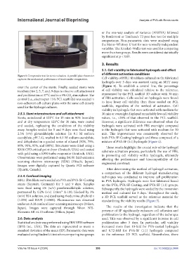
Figure 3. Compression test for stent evaluation. A parallel plate rheometer
captures the mechanical performance of stents under compression. Cell viability of HFL1 fibroblasts cultured on the fabricated
hydrogels over 3 days was assessed using an MTT assay
over the center of the stents. Finally, seeded stents were (Figure 4). To establish a control line, the percentage
incubated for 2, 5, 7, and, 9 days to observe cell attachment of cell viability was calculated relative to the reference,
and proliferation at 37°C and in a 5% CO atmosphere. The represented by the PCL scaffold 3D culture with 30 min
2
control (i.e., electrospun 15% PCL scaffolds) was seeded in of FBS activation. Cells seeded on hydrogels were found
non-adherent cell culture plates with the same cell density to have lower cell viability than those seeded on PCL
used for the hydrogel cultures. scaffolds, regardless of the method of activation. Cell
viability in hydrogels that were activated with medium for
2.5.3. Stent microstructure and cell attachment 30 min or overnight displayed remarkably low cell viability
Stents, autoclaved at 121°C for 15 min in 90% humidity values, i.e., <20% of that observed in the PCL scaffold.
and at dry temperature 121°C for 30 min, were coated However, a significant difference was observed when the
and seeded, replicating the conditions of the viability hydrogels were activated with FBS overnight compared
assay. Samples seeded for 5 and 9 days were fixed using to the hydrogels that were activated with medium for 30
2.5% (v/v) glutaraldehyde solution (in 0.1 M sodium min. This improvement was consistently observed for
cacodylate, pH 7.4), washed in 0.1 M sodium cacodylate, both PVA-SF-Coating hydrogels and those composed of a
and dehydrated in a graded series of ethanol (50%, 75%, mixture of PVA-SF (1:1) hydrogels (Figure 4).
80%, 90%, 95%, and 100%). Structures were dried using a These results highlight the crucial role of the hydrogel
K850 CPD critical point dryer (Emitech, USA) and coated substrate activation process, particularly the use of FBS,
with gold using a K950 turbo evaporator (Emitech, USA). in promoting cell viability within hydrogels, ultimately
Observations were performed using S4100 field emission affecting the performance and biocompatibility of the
scanning electron microscopy (SEM) (Hitachi, Japan). engineered construct.
Images were digitally captured by Quartz PCI software
(Quartz, Canada). After determining the method of hydrogel activation,
a comparison of the different hydrogel manufacturing
2.5.4. Confocal imaging techniques was conducted to improve cell proliferation
HFL1 fibroblasts were seeded on PVA and PVA-SF-Coating in PVA hydrogels. Hydrogels were first fabricated based
stents (Sarstedt, Germany) for 5 and 9 days. Samples on the PVA, PVA-SF-Coating, and PVA-SF (1:1) groups.
were fixed using 4% (w/v) paraformaldehyde solution, Subsequently, the hydrogels were seeded by the immersion
permeated by 0.2% (v/v) Triton™ X-100, blocked by 3% method and cultured for 3 days. Throughout the study,
(w/v) BSA solution, and dyed using rhodamine-phalloidin a 3D PCL scaffold served as the reference material for
(1:250) and DAPI (1:1000). Fluorescence was observed standardizing the viability results (Figure 5).
under an A1R confocal laser scanning microscope (Nikon,
Japan). Images were captured through Nikon NIS- The results of the investigation indicate that the
Elements AR v4.10 software (Nikon, Japan). presence of SF significantly enhances cell adhesion and
proliferation to the hydrogel, regardless of the technique
2.6. Data analysis used. This was observed by a significant increase in cell
Statistical analysis was performed using IBM SPSS software proliferation after 3 days. In particular, cell viability
(SPSS Inc., USA). The data are represented as mean ± increased more than 10-fold for PVA-coated hydrogels
standard deviation of the mean (SD). Parametric data were and 8.72-fold for PVA-SF (1:1) hydrogels compared
evaluated using Student’s t-test when comparing two groups to the reference 3D PCL scaffold. Nevertheless, there
Volume 10 Issue 4 (2024) 286 doi: 10.36922/ijb.3444