
Page 336 - IJB-10-4
P. 336
International Journal of Bioprinting 3D bioprinting of composite hydrogels
For a bioink, the apparent viscosity should be both Therefore, 2.5% GG was selected for further study.
low enough to easily extrude and high enough to finely Interestingly, with the addition of 3PEI, 4PEI, and 5PEI,
maintain the structure of sequential layers. 81,82 From the light transmittance increased to 88.71% ± 1.49%,
Figure 4c, it is apparent that all bioinks were shear thinning, 87.70% ± 1.29%, and 87.70% ± 1.11%, respectively, in
as the viscosity decreased with increasing shear rate. The comparison with the 2.5GG hydrogel (Figure 5b). After
viscosity value for pure GG at a low shear rate of 1.08 s 3D bioprinting the scaffolds, no substantial difference
−1
was lower (117 ± 2.11 Pa·s) than GG–CA (299 ± 2.23 Pa·s) was observed in the transparency of the composites
and GG–3PEI (172 ± 1.71 Pa·s) due to the greater number (85.31% ± 2.19% for 2.5GG, 89.33% ± 1.84% for GG–
of free OH groups in the GG structure. In addition, the 3PEI, 89.33% ± 1.49% for GG–4PEI, and 86.89% ±
in-situ gelling capacity of the bioinks was qualitatively 2.49% for GG–5PEI) (Figure 5c). A complete statistical
assessed. As displayed in Figure 4d, the GG–CA hydrogel comparison of transparency corresponding to the various
formed a gel after 15 min, while the gelation of GG–CA bioink formulations is presented in Table S2, Supporting
mixed with PEI required 30 min. This could be due to a Information. These values are close to those of native
84
79
decrease in the dispersion viscosity in the presence of PEI corneal tissue (87%). Ren et al. reported values of
that hampers crosslinking and gelation of GG by CA. 78% and 60% for the transparency of pure collagen
and collagen-polycaprolactone corneal membranes,
The 3D model of a cylinder had outer and inner respectively. Hosseinian et al. developed methacrylated
85
diameters of 10 and 5 mm, respectively, and consisted of gelatin (GelMA) and corneal dECM-containing
12 layers (Figure 4e). Printed structures were compared to constructs that exhibited a significant increase in light
this model structure to calculate the error and shape fidelity transmittance to 53.6% compared to that of corneal
(Table 3). The GG–CA and GG–3PEI composite hydrogels dECM (5.84%). Hasirci et al. reported transparency
86
displayed superior shape fidelity and structural integrity values of 70–75% for cell-laden GelMA and poly(2-
compared to pure GG. The GG–3PEI structure had a 2.1% hydroxyethyl methacrylate) (pHEMA) interpenetrating
(outer diameter) and 16% (inner diameter) deviation from network hydrogels as a corneal stroma replacement. In
the model. The viscosity and the shape fidelity ratio of GG– another study, silk–gelatin composite scaffolds fabricated
3PEI were similar to GG–CA. Therefore, the presence of by electrospinning permitted 78% light transmittance.
83
PEI had no negative effect on printability and shape fidelity Thus, the GG–PEI composite scaffolds described here
when used in 3D bioprinting. display greater light transmittance than other biomaterials
designed for use in corneal tissue engineering.
3.4. Transparency
Transparency is a key parameter in corneal tissue 3.5. In vitro degradation
engineering, essential for fabricating corneal scaffolds. The in vitro degradation profile of the scaffolds was
83
The transparency of GG makes it a potential candidate investigated by immersing the scaffolds in PBS for 14 days.
for corneal tissue engineering. The percentage of light As observed in Figure 6, pure GG scaffolds cross-linked
transmittance of the samples was measured in the range with CA only degraded the least (19.68% ± 1.10% on day
of UV–visible spectrum, as displayed in Figure 5. The 3, 33.01% ± 1.22% on day 7, and 36.26% ± 2.0% on day
transparency of the 2.5GG and 3GG hydrogels was 82.41% 14), compared to scaffolds that were also functionalized
± 2.10% and 76.91% ± 2.29%, respectively, suggesting with 3% PEI (29.63% ± 1.11% on day 3, 34.66% ± 1.32% on
that increased GG concentration attenuated visible day 7, and 48% ± 2.14% on day 14). The gel content had a
light transmittance through the material (Figure 5a). considerable influence on the degradation of the hydrogels,
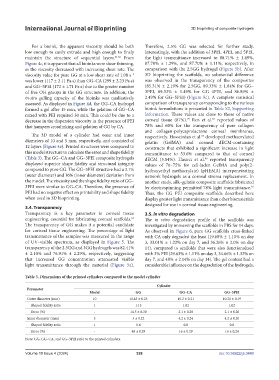
Table 3. Dimensions of the printed cylinders compared to the model cylinder
Cylinder
Parameter
Model GG GG–CA GG–3PEI
Outer diameter (mm) 10 11.45 ± 0.23 10.2 ± 0.21 10.21 ± 0.19
Shaped fidelity ratio 1 1.15 1.02 1.02
Error (%) - 14.5 ± 0.30 2.1 ± 0.28 2.1 ± 0.26
Inner diameter (mm) 5 3 ± 0.22 4.2 ± 0.24 4.2 ± 0.20
Shaped fidelity ratio 1 0.6 0.8 0.8
Error (%) - 40 ± 0.29 16 ± 0.29 16 ± 0.24
Note: GG, GG–CA, and GG–3PEI refer to the printed cylinders.
Volume 10 Issue 4 (2024) 328 doi: 10.36922/ijb.3440