
Page 339 - IJB-10-4
P. 339
International Journal of Bioprinting 3D bioprinting of composite hydrogels
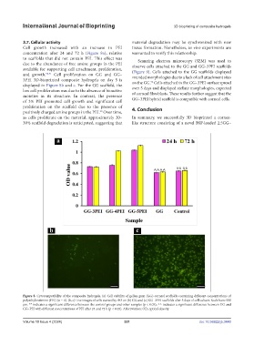
3.7. Cellular activity material degradation may be synchronized with new
Cell growth increased with an increase in PEI tissue formation. Nonetheless, in vivo experiments are
concentration after 24 and 72 h (Figure 8a), relative warranted to verify this relationship.
to scaffolds that did not contain PEI. This effect was Scanning electron microscopy (SEM) was used to
due to the abundance of free amine groups in the PEI observe cells attached to the GG and GG–3PEI scaffolds
available for supporting cell attachment, proliferation, (Figure 9). Cells attached to the GG scaffolds displayed
and growth. 90,91 Cell proliferation on GG and GG– rounded morphologies due to a lack of cell attachment sites
3PEI 3D-bioprinted composite hydrogels on day 5 is on the GG. Cells attached to the GG–3PEI surface spread
92
displayed in Figure 8b and c. For the GG scaffold, the
low cell proliferation was due to the absence of bioactive over 5 days and displayed stellate morphologies, expected
moieties in its structure. In contrast, the presence of corneal fibroblasts. These results further suggest that the
of 3% PEI promoted cell growth and significant cell GG–3PEI hybrid scaffold is compatible with corneal cells.
proliferation on the scaffold due to the presence of
positively charged amine groups in the PEI. Over time, 4. Conclusion
20
as cells proliferate on the material, approximately 20– In summary, we successfully 3D bioprinted a cornea-
30% scaffold degradation is anticipated, suggesting that like structure consisting of a novel BSP-loaded 2.5GG–
Figure 8. Cytocompatibility of the composite hydrogels. (a) Cell viability of gellan gum (GG) corneal scaffolds containing different concentrations of
polyethyleneimine (PEI) (n = 4). (b, c) Live images of cells stained by AO on (b) GG and (c) GG–3PEI scaffolds after 5 days of cell culture. Scale bars: 500
µm. ** indicates a significant difference between the control groups and other samples (p < 0.05); ^^ indicates a significant difference between GG and
GG–PEI with different concentrations of PEI after 24 and 72 h (p < 0.05). Abbreviation: OD, optical density.
Volume 10 Issue 4 (2024) 331 doi: 10.36922/ijb.3440