Page 470 - IJB-10-4
P. 470
International Journal of Bioprinting                                  3D-bioprinted peripheral nerve scaffold


            2.1.3. Shear rate-viscosity and                    (M8211; Solarbio, China) and 1% penicillin/streptomycin
            temperature-viscosity tests                        (15140-122;  Gibco,  USA),  without  fetal  bovine  serum
            The Discovery HR2 rotational rheometer was also utilized   (FBS) (10099–141; Gibco, USA) for a duration of 24 h.
            to  assess  the  viscosity of the  hydrogel. By  subjecting   For the following 72 h, the medium was changed to the
            the bioink to a 1% strain for 200 s at 20℃, we obtained   standard culture medium (αMEM containing 20% FBS
            composite viscosity–shear rate curves. Subsequently,   and 1% penicillin/streptomycin) supplemented with 35
            a shear rate of 0.1–1000 s  was used to establish the   ng/ml all-trans-retinoic acid (A8539; ApexBio, USA).
                                   -1
            correlation between the shear rate and viscosity of the   After 3 days, the medium was switched to standard culture
            hydrogel. To obtain the temperature–viscosity curve, 1 Hz   medium supplemented with 5 μM forskolin (B1421;
            frequency and 1% strain were applied to the hydrogel for   ApexBio, USA), 5 ng/ml platelet-derived growth factor AA
            245 s at varying temperatures (10–42℃).            (HZ-1215; Proteintech, USA), 10 ng/ml basic fibroblast
                                                               growth factor (HZ-1285; Proteintech, USA), and 200 ng/ml
            2.1.4. Tensile modulus test and maximum tensile test  human neuregulin-β1 (ab50227; Abcam, USA). Thereafter,
            A tensile meter (SH-10; WD, China) was used to measure the   the medium was renewed every 3 days, and the SHEDs
            maximum tensile force of the hydrogel (after crosslinking),   were  maintained  at  least  2 weeks  before examination  or
            PCL, and composite scaffolds. The constructed scaffolds
            (from section 2.3. 3D bioprinting) would be hooked to   bioprinting. Flow cytometry analysis was performed to
            both ends of the tensile meter and stretched at a rate of   evaluate the expression levels of the surface antigens CD29,
            0.5 mm/s until fracture. The instrument would record the   CD34, CD45, CD73, CD90, and CD105.
            maximum tensile force endured during the stretch.  2.3. 3D bioprinting
            2.1.5. Scanning electron microscopy                Briefly, a cuboid (with 10 mm length, 10 mm width, and
            The hydrogels were freeze-dried and gold-coated prior to   0.3 mm height) was designed using a 3D bioprinter (BMP-
            observing the micromorphology using a scanning electron   C300-T300-IN3; Medprin, China). The cuboid was designed
            microscope (SEM) (SU8020; Hitachi, Japan). The Image   in two types: linear or reticulated cuboid. Thereafter, PCL
            J software was used for porosity measurements, and the   was placed in the high-temperature melting cylinder
            average porosity was obtained.                     (Cylinder 1), while 1 ml injector was placed into Cylinder 2
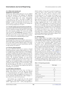
                                                               after mixing the scSHEDs with the hydrogel. A 3D-printing
            2.1.6. Fourier infrared analysis                   pinhead, with an internal diameter of 100 μm, was selected
            We used a NICOLET IS10 Fourier transform infrared   for Cylinder 1, while another pinhead, with a diameter of
            (FTIR) spectrometer (Thermo Fisher Scientific Inc., USA)   160 μm, was selected for Cylinder 2.  Table 1 displays the
            to investigate the chemical composition of the hydrogels.   3D-bioprinting parameters. For the linear cuboid (Figure 3I),
            To clarify the stability of the hydrogel components after the   Cylinder 1 was used to print the first layer, while Cylinder 2
            introduction of the RGD peptide, we performed assays on   printed the second layer. As for the reticulated cuboid, both
            two hydrogels: 6% RGD-Alg/5% GelMA and 6% Alg/5%   layers were printed by Cylinder 2, and the cuboid was used
            GelMA. The lyophilized hydrogel was meticulously   for subsequent cell Live/Dead, immunofluorescence, and
            pulverized in an agate mortar with 200 mg of KBr at a ratio   cytoskeleton staining purposes. After printing the construct,
            of roughly 1:20 and compacted into a slender section. The   it was solidified via 430 nm blue light exposure for 10 s, and
            sample was then positioned within an automated sample
            enclosure for approximately 100 s for identification, while   Table 1. 3D-bioprinting parameters
            the spectral range was 400–4000 cm  and the resolution
                                          -1
            was 4 cm . The FTIR spectra were analyzed after baseline   Parameter             Value/ranges
                   -1
            and background corrections.                         Temperature (℃)
            2.2. Cell culture and induction                     Cylinder 1                       90
            SHEDs were offered by  the Oral  Stem Cell Bank  of   Cylinder 2                    20–25
            Beijing, Tason Biotech Co., Ltd. The cells were cultured   Nozzle 1                  100
            in a mesenchymal stem cell medium (MSCM) (#7501;    Ambient                          20
            ScienCell, USA) within a humidified environment at 37℃,   Fill density (%)           60
            and the cells were subcultured every 3 days. Passages 4–7   XY plotting speed (mm/s)  50
            were collected for experimental purposes.
                                                                Inner diameter (μm)
               Cell induction was initiated when the cells reached   Nozzle 1                    100
            a confluency of ≥ 80%, and the medium was changed to   Nozzle 2                      220
            αMEM  supplemented  with  1  mM  β-mercaptoethanol

            Volume 10 Issue 4 (2024)                       462                                doi: 10.36922/ijb.2908

