
Page 477 - IJB-10-4
P. 477
International Journal of Bioprinting 3D-bioprinted peripheral nerve scaffold
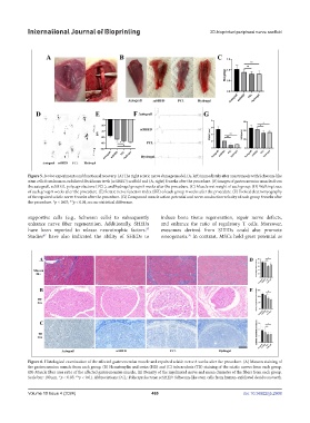
Figure 5. In vivo experiments and functional recovery. (A) The right sciatic nerve damage model, (A, left) immediately after anastomosis with Schwann-like
stem cells from human-exfoliated deciduous teeth (scSHED) scaffold and (A, right) 8 weeks after the procedure. (B) Images of gastrocnemius muscles from
the autograft, scSHED, polycaprolactone (PCL), and hydrogel groups 8 weeks after the procedure. (C) Muscle wet weight of each group. (D) Walking trace
of each group 8 weeks after the procedure. (E) Sciatic nerve function index (SFI) of each group 8 weeks after the procedure. (F) Evoked electromyography
of the repaired sciatic nerve 8 weeks after the procedure. (G) Compound muscle action potential and nerve conduction velocity of each group 8 weeks after
the procedure. *p < 0.05; **p < 0.01; ns: no statistical difference.
supportive cells (e.g., Schwann cells) to subsequently induce bone tissue regeneration, repair nerve defects,
enhance nerve fiber regeneration. Additionally, SHEDs and enhance the ratio of regulatory T cells. Moreover,
have been reported to release neurotrophic factors. exosomes derived from SHEDs could also promote
37
Studies have also indicated the ability of SHEDs to osteogenesis. In contrast, MSCs hold great potential as
36
47
Figure 6. Histological examination of the affected gastrocnemius muscle and repaired sciatic nerve 8 weeks after the procedure. (A) Masson staining of
the gastrocnemius muscle from each group. (B) Hematoxylin and eosin (HE) and (C) tuberculosis (TB) staining of the sciatic nerves from each group.
(D) Muscle fiber area ratio of the affected gastrocnemius muscle. (E) Density of the myelinated nerve and mean diameter of the fibers from each group.
Scale bar: 100 μm. *p < 0.05; **p < 0.01. Abbreviations: PCL: Polycaprolactone; scSHED: Schwann-like stem cells from human-exfoliated deciduous teeth.
Volume 10 Issue 4 (2024) 469 doi: 10.36922/ijb.2908