
Page 102 - IJB-5-1
P. 102
Near-field electrospinning of a polymer/bioactive glass composite to fabricate 3D biomimetic structures
been to combine the electrospinning and 3D printing ultrasonication and materials were uniformly mixed for
techniques to provide a nanofiber mesh in between 5 min at 2000 RPM in a planetary mixer (SpeedMixer™,
the 3D printed macroporous layers . An alternative FlakTek Inc., Landrum, SC). The weight ratio of
[8]
approach to the traditional electrospinning is called materials was selected in such a way to provide a 20%
near-field electrospinning (NFES), where the substrate glass in weight in the fabricated composite scaffolds
distance from nozzle tip is decreased to control the fiber after eventual CF evaporation.
deposition [9,10] . In NFES technique, fiber instability is
restricted because of the shorter substrate distance and 2.2. Scaffold Fabrication and Characterization
deposition is precisely controlled to obtain the desired For NFES, a home-built, three-axis gantry system with
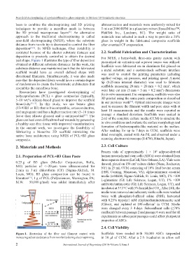
part shape. Figure 1 illustrates the type of fiber deposition pressurized air extrusion and a power source was utilized.
obtained at different substrate distances. In this work, the Scaffolds were fabricated on an aluminum foil placed on
substrate distance was maintained such that the fabricated a metal substrate and a custom-made software interface
scaffold would have an overall defined shape with was used to control the printing parameters including
directional filaments. Simultaneously, it was also made applied voltage, air pressure, and printing speed. A metal
sure that the deposited fibers would have a certain degree tip (0.25 mm internal diameter) was used to fabricate
of randomness to create the biomimetic architecture that scaffolds measuring 20 mm × 20 mm × 0.2 mm , which
3
resembles the cancellous bone. were later cut into (5 mm × 5 cm × 0.2 mm ) dimensions
3
Researchers have investigated electrospinning of
polycaprolactone (PCL) + glass composites (typically, for in vitro assessment. For comparison, scaffolds were also
5–10 wt.% silicate-based glass) to improve the scaffold 3D printed using the same paste composition as described
[14]
bioactivity [11,12] . In this study, we use borate glass in our previous work . Optical microscopic images were
(13-93B3 or B3) that is biocompatible, osteoconductive, used to measure the filament width and pore sizes with at
and angiogenic and has a higher reaction rate (5–10 times least 10 measurements and the results were reported as
faster than silicate glasses) and is antimicrobial . The average ± standard deviation. Scaffolds were soaked in
[13]
glass can heal even difficult-to-heal wounds by generating 2 ml of the complete culture media (CCM) to simulate the
a healthy scar-free tissue with improved vascularization. in vitro conditions and evaluate the surface morphology and
In the current work, we investigate the feasibility of formation of hydroxyapatite-like material on the surface.
fabricating a bioactive 3D scaffold mimicking the After soaking for up to 7 days in CCM, scaffolds were
native bone architecture using NFES of PCL+B3 glass dried overnight, coated with Au-Pd, and observed under a
composite. scanning electron microscope (S-4700, Hitachi, Japan).
2. Materials and Methods 2.3. Cell Culture
Frozen vials of approximately 1 × 10 adipose-derived
6
2.1. Preparation of PCL+B3 Glass Paste human mesenchymal stem cells (ASCs) were obtained from
three separate donors (LaCell, New Orleans, LA). Vials were
0.25 g of B3 glass (Mo-Sci Corporation, Rolla, thawed, plated on 150 cm culture dishes (Nunc, Rochester,
2
MO) particles of <~20 µm were ultrasonicated for NY) in 25 mL CCM consisting of 10% fetal bovine serum
2 min in 3 ml chloroform (CF) (Sigma-Aldrich, St. (FBS, Corning, Manassas, VA), alpha-minimum essential
Louis, MO). B3 glass composition can be found in media (α-MEM, Sigma-Aldrich, St. Louis, MO), 1% ×100
literature , 1 g of PCL (Polysciences, Warrington, PA; L-glutamine (GE Life Sciences, Logan, UT), 1% ×100
[14]
M.W. – 50,000 g/mol) was added immediately after
antibiotic/antimycotic (GE Life Sciences, Logan, UT), and
incubated at 37.5°C with 5% humidified CO . After 24 h, the
2
media were removed and adherent, viable cells were washed
twice with phosphate-buffered saline (PBS), harvested
with 0.25% trypsin/1 mM ethylenediaminetetraacetic acid
(Gibco), and replated at 100 cells/cm in CCM. Media
2
were changed every 3–4 days. Subconfluent cells (≤70%
confluent) between passage 2 and passage 6 were used for all
experiments as subsequent passages could affect pluripotent
properties of ASCs.
2.4. Cell Viability
Figure 1. Illustration of the fiber and filament control with Scaffolds were seeded with 30,000 ASCs suspended
increasing substrate distance from nozzle tip during electrospinning. in 30 µl of CCM. After a 2 h incubation to allow cell
2 International Journal of Bioprinting (2019)–Volume 5, Issue 1