
Page 105 - IJB-5-1
P. 105
Kolan KCR, et al.
a b c d
e f g h
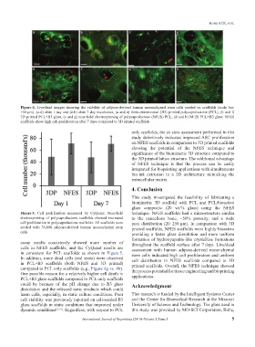
Figure 4. Live/dead images showing the viability of adipose-derived human mesenchymal stem cells seeded on scaffolds (scale bar:
100 µm). (a-d) after 1 day and (e-h) after 7-day incubation, (a and e) three-dimensional (3D) printed polycaprolactone (PCL), (b and f)
3D printed PCL+B3 glass, (c and g) near-field electrospinning of polycaprolactone (NFES) PCL, (d and h) NFES PCL+B3 glass. NFES
scaffolds show high cell proliferation after 7 days compared to 3D printed scaffolds
only scaffolds, the in vitro assessment performed in this
study definitively indicates improved ASC proliferation
on NFES scaffolds in comparison to 3D printed scaffolds
showing the potential of the NFES technique and
significance of the biomimetic 3D structure compared to
the 3D printed lattice structure. The additional advantage
of NFES technique is that the process can be easily
integrated for bioprinting applications with simultaneous
bio-ink extrusion in a 3D architecture mimicking the
extracellular matrix.
4. Conclusion
This study investigated the feasibility of fabricating a
biomimetic 3D scaffold with PCL and PCL/bioactive
glass composite (20 wt.% glass) using the NFES
Figure 5. Cell proliferation measured by CyQuant. Near-field technique. NFES scaffolds had a microstructure similar
electrospinning of polycaprolactone scaffolds showed increased to the cancellous bone, ~50% porosity, and a wide
cell proliferation in polycaprolactone scaffolds. All scaffolds were pore distribution (20–250 µm). In comparison with 3D
seeded with 30,000 adipose-derived human mesenchymal stem printed scaffolds, NFES scaffolds were highly bioactive
cells providing a faster glass dissolution and more uniform
formation of hydroxyapatite-like crystalline formations
assay results consistently showed more number of throughout the scaffold surface after 7 days. Live/dead
cells in NFES scaffolds, and the CyQuant results are assessment with human adipose-derived mesenchymal
in consistent for PCL scaffolds as shown in Figure 5. stem cells indicated high cell proliferation and uniform
In addition, more dead cells (red spots) were observed cell distribution in NFES scaffolds compared to 3D
in PCL+B3 scaffolds (both NFES and 3D printed) printed scaffolds. Overall, the NFES technique showed
compared to PCL only scaffolds (e.g., Figure 4g vs. 4h). the process potential for tissue engineering and bioprinting
One possible reason for a relatively higher cell death in
PCL+B3 glass scaffolds compared to PCL only scaffolds applications.
could be because of the pH change due to B3 glass Acknowledgment
dissolution and the released ionic products which could
harm cells, especially, in static culture conditions. Poor This research is funded by the Intelligent Systems Center
cell viability was previously reported on cell-seeded B3 and the Center for Biomedical Research at the Missouri
glass scaffolds in static conditions that improved under University of Science and Technology. The glass used in
dynamic conditions [2,17] . Regardless, with respect to PCL this study was provided by MO-SCI Corporation, Rolla,
International Journal of Bioprinting (2019)–Volume 5, Issue 1 5