
Page 47 - IJB-5-1
P. 47
Cheptsov VS, et al.
infections (e.g., wounds, oral cavity, and cystic fibrosis
of the lungs) where tissues are often colonized by several
species of bacteria simultaneously. The true power of 3D
cell printing lies in the ability to organize the microbial
communities in the unlimited range of geometries.
Micro-3D cell printing can also be a valuable tool for
the investigations of mechanisms and dynamics of the
adaptive responses to environmental conditions.
3. Laser Bioprinting
Over the past two decades, bioprinting, including the
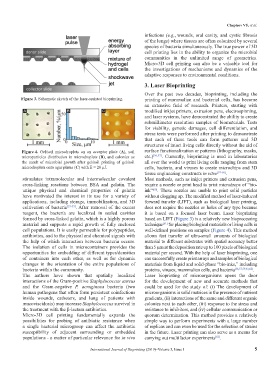
Figure 3. Schematic sketch of the laser-assisted bioprinting. printing of mammalian and bacterial cells, has become
an extensive field of research. Printers, starting with
A B C modified inkjet printers, extrusion pens, electrospinning,
and laser systems, have demonstrated the ability to create
submillimeter resolution samples of biomaterials. Tests
for viability, genetic damages, cell differentiation, and
stress tests were performed after printing to demonstrate
that each of these tools can form patterns and 3D
structures of intact living cells directly without the aid of
Figure 4. Gel/soil microdroplets on an acceptor plate (A), soil surface functionalization or patterns (lithography, masks,
microparticles distribution in microdroplets (B), and colonies as etc.) [56,57] . Currently, bioprinting is used in laboratories
the result of microbial growth after gel/soil printing of gel/soil all over the world to print living cells ranging from stem
microdroplets onto agar plates (C) with E = 20 µJ. cells, bacteria, and viruses to create microchips and 3D
tissue engineering constructs in vitro [57-60] .
stimulates intramolecular and intermolecular covalent Most methods, such as inkjet printers and extrusion pens,
cross-linking reactions between BSA and gelatin. The require a nozzle or print head to print microdrops of “bio-
unique physical and chemical properties of gelatin ink” . These nozzles are unable to print solid particles
[60]
have motivated the interest in its use for a variety of without clogging up. The modified method of laser-induced
applications, including storage, immobilization, and 3D forward transfer (LIFT), such as biological laser printing,
cultivation of bacteria [53-55] . After removal of the excess does not require the nozzles or holes of any type because
reagent, the bacteria are localized in sealed cavities it is based on a focused laser beam. Laser bioprinting
formed by cross-linked gelatin, which is a highly porous based on LIFT (Figure 3) is a relatively new bioprocessing
material and supports a rapid growth of fully enclosed technique for placing biological materials or living cells in
[61]
cell populations. It is easily permeable for polypeptides, well-defined positions on samples (Figure 4). This method
antibiotics, and to the physical and chemical signals with allows fast transfer of ultra-small amounts of biological
the help of which interaction between bacteria occurs. material to different substrates with spatial accuracy better
The isolation of cells in microcontainers provides the than 5 µm at the deposition rate up to 100 pixels of biological
opportunities for embedding of different types/densities material per second. With the help of laser bioprinting, one
of containers into each other, as well as for dynamic can successfully create print arrays and samples of biological
changes in the orientation of the entire populations of materials from liquid and solid-phase “bio-inks,” including
bacteria within the community. proteins, viruses, mammalian cells, and bacteria [56,57,59,61,62] .
The authors have shown that spatially localized Laser bioprinting of microorganisms opens the door
interactions of the Gram-positive Staphylococcus aureus for the development of new and accurate methods that
and the Gram-negative P. aeruginosa bacteria (two could be used for the study of: (i) The development of
human pathogens that often form persistent coinfections microorganisms in solid matrices in the presence of nutrient
inside wounds, catheters, and lung of patients with gradients, (ii) interactions of the same and different organic
mucoviscidosis) may increase Staphylococcus survival in colonies next to each other, (iii) response to the stress and
the treatment with the β-lactam antibiotics. resistance to inhibitors, and (iv) cellular communication or
Micro-3D cell printing fundamentally expands the quorum determination. This method provides a relatively
possibilities for probing of antibiotic resistance when simple way to perform experiments with a large number
a single bacterial microgroup can affect the antibiotic of replicas and can even be used for the selection of strains
susceptibility of adjacent surrounding or embedded in the future. Laser printing can also serve as a means for
populations - a matter of particular relevance for in vivo carrying out multifactor experiments .
[63]
International Journal of Bioprinting (2019)–Volume 5, Issue 1 5