
Page 48 - IJB-5-1
P. 48
New microorganism isolation techniques with emphasis on laser printing
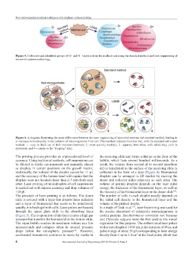
Figure 5. Cultivated and identified groups of G+ and G− bacteria from the mollisol soil using the standard method and laser engineering of
microbial systems technology.
Figure 6. A diagram illustrating the main differences between the laser engineering of microbial systems and standard method, leading to
an increase in biodiversity in the isolation of microorganisms from soil. The numbers indicate microbes that, with the standard cultivation
method: 1 - easy to flush out of their microenvironment, 2 - most actively multiply, 3 - separate from those with which they exist in
symbiosis, and 4 - remain in the “sleeping” state.
The printing process provides an unprecedented level of the receiving slide and forms a thin jet at the front of the
accuracy. Using traditional methods, cell suspensions can bubble, which lasts several hundred milliseconds. As a
be diluted in sterile environments and manually placed result, the volume from several pl to several nanoliters
as droplets in certain positions on the growth matrix; (nl) is transferred to the surface of the receiving slide (a
realistically, the volume of the droplet cannot be <1 µl, collector) in the form of a drop (Figure 4). Biomaterial
and the accuracy of the human hand will require that the droplets can be arranged in 2D models by moving the
droplets were not located closer than 2–3 mm from each donor and collector slides relatively to each other. The
other. Laser printing of microdroplets of cell suspensions volume of printed droplets depends on the laser pulse
is carried out with micron accuracy and drop volumes of energy, the thickness of the biomaterial layer, as well as
<10 pl. the viscosity of the biomaterial layer on the donor slide .
[65]
The principle of laser printing is as follows: The donor The number of cells in each droplet usually depends on
slide is covered with a layer that absorbs laser radiation the initial cell density in the biomaterial layer and the
and a layer of biomaterial that needs to be transferred; volume of the printed droplet.
usually, it is hydrogel with cells. Laser pulses are focused In a study of Taidi et al. , laser bioprinting was used for
[63]
through the upper glass slide in the absorbing layer the precise placement of eukaryotic microorganisms in
(Figure 3). The evaporation of this layer creates a high gas certain patterns. Saccharomyces cerevisiae var. bayanus
pressure that transfers the biomaterial to the bottom slide. and Chlorella vulgaris were the first used as the model
The vapor bubble reaches its maximum volume in a few organisms for this purpose. The authors used laser pulses
microseconds and collapses when its internal pressure with a wavelength of 1064 nm, pulse duration of 10 ns, and
drops below the atmospheric pressure . However, pulse energy of about 20 μJ corresponding to laser energy
[64]
accelerated biomaterial continues to move by inertia to density from 1 up to 2 J/cm at the focal point, which was
2
6 International Journal of Bioprinting (2019)–Volume 5, Issue 1