
Page 32 - IJB-5-2
P. 32
A novel inkjet system for live cell bioprinting
addition to the requirement for the preparation of ribbons 2. Materials and Methods
of cells and hydrogels. Conversely, inkjet printing, and
more generally, droplet-based bioprinting , have great 2.1. Cell Cultures
[5]
promise as a simple and efficient method for the precise
patterning of multiple cell types and bioink components All cells were cultured in a 5% CO incubator at 37.0°C
2
including active biomacromolecules , especially since and passaged manually every 2 to 3 days to maintain a
[6]
a drop-on-demand control of small volumes down to a subconfluent state. NIH/3T3 mouse fibroblast cell line
few hundred picoliters can be expected. However, inkjet (clone 5611, JCRB Cell Bank) and normal human dermal
technology has several limitations that impair its further fibroblasts (NHDF, CC-2509, Lonza Inc.) were cultured
adoption in 3D construction. Although some of the earliest in Dulbecco’s Modified Eagle’s Medium (Thermo
reports of successful bioprinting in the mid-2000s were Fisher Scientific Inc.) supplemented with 10% fetal
inkjet based [7-9] , few concrete results of fully functional bovine serum (Biowest) and 1% penicillin-streptomycin
inkjet-produced tissues have been reported to date. (26253-84, NACALAI TESQUE, INC). Human umbilical
The first notable limitation of inkjet bioprinting is blood vein endothelial cells (HUVEC, CC-2519, Lonza
that ejecting large cell-sized particles from common Inc.) were cultured in endothelial cell growth medium
printheads is a challenge. Successful ejection has been (EGM, Lonza Inc.) with supplements as recommended
reported [10-13] , and acoustic ejection achieved in live by the manufacturer. For bioink preparation, the cells
cell printing ; however, cell sedimentation inside the were washed twice with Dulbecco’s phosphate-buffered
[13]
printhead chamber and clogging of the nozzle is expected saline without calcium and magnesium (DPBS, Thermo
to rapidly compromise any reliable control of droplet Fisher Scientific Inc.), detached with 0.05% Trypsin-
formation over the length of time required to produce a EDTA (25300054, Thermo Fisher Scientific Inc.), and
3D tissue. Second, the range of materials that can be used centrifuged at 400 g for 5 min at 4°C. The cell pellets
as substrates to carry the cells is limited to ejectable low- were re-suspended in fresh DPBS at room temperature
viscosity liquids so that shaping fine 3D structures with and used within 30 min after suspension.
suitable mechanical properties is particularly challenging.
Various strategies have been reported including coprinting 2.2. Inkjet Print Head Development
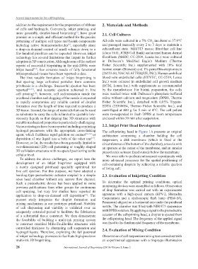
hydrogel precursors with the appropriate cross-linking The cell-printing head in Figure 1A presents an original
agent, which facilitates rapid gelation on contact [14-16] or architecture comprising a chamber holding the cell
deposition of one liquid into a bath of the other one . suspension, a disk membrane (which is fixed at the
[17]
However, so far, the results have been generally limited to circumference of the bottom of the chamber), a nozzle with
two-dimensional (2D) cell patterning or roughly shaped an aperture at the center of the membrane, and an annular
3D cell-laden structures with no spatial positioning at the piezoelectric actuator fixed outside below the membrane
cellular level. We were able to perform subsequent experiments with
To address the above challenges, we report here the more advanced processes for the spatial positioning of
development of an inkjet bioprinter equipped with cell-containing droplets by achieving a reliable ejection
a newly designed printhead specially optimized for of living cell.
live cell ejection. For this purpose, we have adapted a
bending-type piezoelectric actuator coupled to a simple 2.3. Evaluation of Inkjetting Condition
open head chamber without any narrow flow channel.
Such a piezoelectric device has been applied in some To determine the optimal printing conditions, optical
previous publications from other groups for continuous monitoring devices were assembled as follows. Observation
cell spraying, but very few studies have reported its of drop formation was carried out with an experimental
application to drop-on-demand cell deposition . The apparatus with a high-speed camera (HPV-2, Shimadzu
[18]
present study integrates the droplet formation and Corporation) and a stroboscopic flash lamp (PE60-SG,
mixing mechanism in our prototype printhead. Stability Panasonic) aligned on a horizontal axis under the printhead
of cell dispensing and viability is validated over an nozzle. The chamber was filled with NIH/3T3 suspension
adequately extended period to facilitate the fabrication with DPBS solution. By applying a signal to the piezoelectric
of a substantial tissue construct. We then demonstrate actuator of the cell-printing head, a droplet is ejected from
the feasibility of building a multi-ink printing system the cell-printing head. The frequency of the applied signal
to construct stratified Mille-Feuille-like structures with was fixed to the fundamental frequency of the membrane.
controlled thickness by alternating cell suspension and 2.4. Evaluation of Mixing Condition
hydrogel layers. Therefore, exploiting the full potential
of inkjet technology promises to facilitate high-precision Observation of cell suspension mixing was carried out with
multi-ink 3D bioprinting. an experimental apparatus with a ring-type illumination
28 International Journal of Bioprinting (2019)–Volume 5, Issue 2