
Page 89 - IJB-5-2
P. 89
Chen Y, et al.
obtained in a multi-well spectrophotometer (Hitachi, coated object (MP), and the PDA/HA-coated object
Tokyo, Japan) at 570 nm with a reference wavelength of (MPHA) are shown in Figure 2. M and MP have no
600 nm. peaks. The peaks of MHA and MPHA at around 2θ=25.7°
and 2θ=31.9° are characteristic of HA precipitates,
2.5 Cell Morphology which occurs during the early mineral phase of bone
[21]
After 12 h of cell culture, the samples with hMSCs were development and fracture healing . The result shows the
washed with cold PBS and fixed by 1.5% glutaraldehyde PDA/HA-coated MED610 object contains a large amount
(Sigma-Aldrich, MO, USA) for 2 h and then were of HA precipitate.
dehydrated by a graded ethanol series for 20 min at each Figure 3 shows the SEM results of MED610 object with
concentration and dried with liquid CO by a critical HA, PDA, or PDA/HA coatings. The PDA/HA coated
2
point dryer device (LADD 28000, LADD, Williston, VT, object presents more HA mineral crystallization. Based
USA). The dried samples were mounted on stubs, coated on these results, it is speculated that the PDA coating
with gold particles, and investigated by SEM (JEOL can effectively assist the bionics of HA mineralization,
JSM-7401F, Tokyo, Japan). thereby producing a hybrid biomaterial having HA. In
addition, the addition of a PDA/HA coating can increase
2.6 Osteogenesis Assay the hardness of the printed object (Figure 4).
After 3 and 7 days of cell culture, the level of 3.2 Cell Proliferation and Morphology
alkaline phosphatase (ALP) activity was evaluated Whether the biomedical materials printed by the 3D
using p-nitrophenyl phosphate (pNPP, Sigma) as the printer can be widely used in the medical field, the
substrate. The samples were mixed with pNPP in 1 M
diethanolamine buffer for 15 min, then stopped by the
addition of 5N NaOH and quantified by absorbance at
405 nm. The experiments were performed in triplicate.
2.7 Statistical Analysis
A one-way variance statistical analysis was used to
evaluate the significance of the differences between
the groups in each experiment. Scheffe’s multiple
comparison test was used to determine the significance of
the deviations in the data for each specimen. In all cases,
the results were considered statistically significant with
P<0.05.
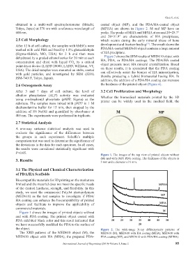
Figure 1. The images of the top view of printed objects without
3. Results (M) and with (MP) PDA coating. The thickness of the objects is
3 mm and a diameter of 6 mm.
3.1 The Physical and Chemical Characterization
of PDA/HA Scaffolds
Biocompatible materials for 3D printing on the market are
limited and the material does not meet the specific needs
of the desired hardness, strength, and flexibility. In this
study, we used the commercial PolyJet photopolymers
(MED610) as the test samples to investigate if PDA/
HA coating can enhance the biocompatibility of printed
objects and facilitate to improve the applicability of
commercial materials.
Figure 1 shows the images of printed objects without
and with PDA coating. The printed object coated with
PDA exhibited black color and this result indicated that
we have successfully modified the PDA to the surface of
the object. Figure 2. The wide-range X-ray diffractometry patterns of
The XRD patterns of the MED610 object (M), the MED610 (M), MED610 with HA coating (MHA), MED610 with
MED610 object with HA (MHA), the prepared PDA- PDA coating (MP), and MED610 with PDA/HA coating (MPHA).
International Journal of Bioprinting (2019)–Volume 5, Issue 2 85