
Page 90 - IJB-5-2
P. 90
The mussel-inspired assisted apatite mineralized on PolyJet material for artificial bone scaffold
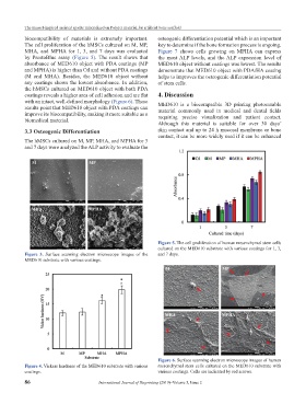
biocompatibility of materials is extremely important. osteogenic differentiation potential which is an important
The cell proliferation of the hMSCs cultured on M, MP, key to determine if the bone formation process is ongoing.
MHA, and MPHA for 1, 3, and 7 days was evaluated Figure 7 shows cells growing on MPHA can express
by PrestoBlue assay (Figure 5). The result shows that the most ALP levels, and the ALP expression level of
absorbance of MED610 object with PDA coatings (MP MED610 object without coatings was lowest. The results
and MPHA) is higher than Ctl and without PDA coatings demonstrate that MED610 object with PDA/HA coating
(M and MHA). Besides, the MED610 object without helps to improves the osteogenic differentiation potential
any coatings shows the lowest absorbance. In addition, of stem cells.
the hMSCs cultured on MED610 object with both PDA
coatings reveals a higher area of cell adhesion and are flat 4. Discussion
with an intact, well-defined morphology (Figure 6). These MED610 is a biocompatible 3D printing photocurable
results point that MED610 object with PDA coatings can material commonly used in medical and dental fields
improve its biocompatibility, making it more suitable as a requiring precise visualization and patient contact.
biomedical material.
Although this material is suitable for over 30 days’
3.3 Osteogenic Differentiation skin contact and up to 24 h mucosal membrane or bone
contact, it can be more widely used if it can be enhanced
The hMSCs cultured on M, MP, MHA, and MPHA for 3
and 7 days were analyzed the ALP activity to evaluate the
Figure 5. The cell proliferation of human mesenchymal stem cells
cultured on the MED610 substrate with various coatings for 1, 3,
Figure 3. Surface scanning electron microscope images of the and 7 days.
MED610 substrate with various coatings.
Figure 6. Surface scanning electron microscope images of human
Figure 4. Vickers hardness of the MED610 substrate with various mesenchymal stem cells cultured on the MED610 substrate with
coatings. various coatings. Cells are indicated by red arrows.
86 International Journal of Bioprinting (2019)–Volume 5, Issue 2