
Page 95 - IJB-5-2
P. 95
Fang A, et al.
However, the research on the glial scar is mainly
based on two-dimensional (2D) culture. Yu et al. [19] used
a sterile plastic pipette to scratch the astrocytes in the 2D
culture to simulate the damage and compared the protein
content to estimate the degree of injury. In addition, they
found that the surrounding part of the damaged area had
also suffered a certain degree of trauma. The response
to the scratching can be mimicked and this kind of 2D
scratch-would model is widely used. Kimura-Kuroda
et al. [20] constructed a glial scar-like structure by 2D
coculturing of meningeal fibroblasts and brain astrocytes
with transforming growth factor-β1 (TGF-β1). The
model can inhibit the neurite outgrowth of neurons
remarkably. In general, the traditional method of 2D cell
culture cannot mimic the cell growth conditions in vivo
and the physiological activities of normal cells. Hence,
it cannot reflect the nature glial scar tissue properly.
However, the three-dimensional (3D) model is expected
to be able to express the glial scar effect from a more
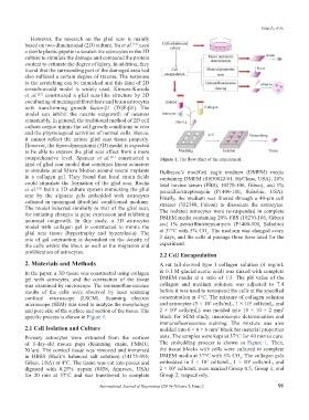
comprehensive level. Spencer et al. [21] constructed a Figure 1. The flow chart of the experiment.
kind of glial scar model that combines linear actuators
to simulate axial Micro Motion around neural implants Dulbecco’s modified eagle medium (DMEM) media
in a collagen gel. They found that local strain fields containing DMEM (SH30022.01, HyClone, USA), 20%
could stimulate the formation of the glial scar. Rocha fetal bovine serum (FBS), 10270-106, Gibco), and 1%
et al. [22] built a 3D culture system mimicking the glial penicillin/streptomycin (P1400-100, Solarbio, USA).
scar by the alginate gels embedded with astrocytes Finally, the medium was filtered through a 40-μm cell
cultured in meningeal fibroblast conditioned medium. strainer (352340, Falcon) to dissociate the astrocytes.
The model behaved similarly to that of the glial scar, The isolated astrocytes were re-suspended in complete
for initiating changes in gene expression and inhibiting DMEM media containing 20% FBS (10270-106, Gibco)
neuronal outgrowth. In this study, a 3D astrocytes and 1% penicillin/streptomycin (P1400-100, Solarbio)
model with collagen gel is constructed to mimic the
glial scar tissue (hypertrophy and hyperplasia). The at 37°C with 5% CO . The medium was changed every
2
rate of gel contraction is dependent on the density of 2 days, and the cells at passage three were used for the
the cells within the block as well as the migration and experiment.
proliferation of astrocytes. 2.2 Cell Encapsulation
2. Materials and Methods A rat tail-derived type I collagen solution (4 mg/mL
In the paper, a 3D tissue was constructed using collagen in 0.1 M glacial acetic acid) was mixed with complete
gel with astrocytes, and the contraction of the tissue DMEM media at a ratio of 1:3. The pH value of the
was examined by microscope. The immunofluorescence collagen and medium solution was adjusted to 7.4
results of the cells were observed by laser scanning before it was used to resuspend the cells at the specified
confocal microscope (LSCM). Scanning electron concentration at 4°C. The mixture of collagen solution
6
5
microscope (SEM) was used to analyze the morphology and astrocytes (5 × 10 cells/mL, 1 × 10 cells/mL, and
6
3
and pore size of the surface and section of the tissue. The 2 × 10 cells/mL) was molded into 10 × 10 × 2 mm
specific process is shown in Figure 1. block for SEM study, macroscopic determination and
immunofluorescence staining. The mixture was also
2.1 Cell Isolation and Culture molded into 6 × 6 × 6 mm block for material properties
3
Primary astrocytes were extracted from the cortices tests. The samples were kept at 37°C for 40 min to cure.
of 1-day-old mouse pups (Kunming strain, FMMU, The embedding process is shown in Figure 1. Then,
Xi’an). The cortical tissue was removed and immersed the tissue blocks with cells were cultured in complete
in HBSS (Hank’s balanced salt solution) (14175-095, DMEM media at 37°C with 5% CO . The collagen gels
2
6
Gibco, USA) at 4°C. The tissue was cut into pieces and embedded in 5 × 10 cells/mL, 1 × 10 cells/mL, and
5
digested with 0.25% trypsin (0458, Amresco, USA) 2 × 10 cells/mL were marked Group 0.5, Group 1, and
6
for 20 min at 37°C and was transferred to complete Group 2, respectively.
International Journal of Bioprinting (2019)–Volume 5, Issue 2 91